17
views
0
recommends
+1 Recommend
0 collections
    0
    shares
      • Record: found
      • Abstract: found
      • Article: found
      Is Open Access

      miR-22 Is a Novel Mediator of Vascular Smooth Muscle Cell Phenotypic Modulation and Neointima Formation

      research-article

      Read this article at

      Bookmark
          There is no author summary for this article yet. Authors can add summaries to their articles on ScienceOpen to make them more accessible to a non-specialist audience.

          Abstract

          Supplemental Digital Content is available in the text.

          Abstract

          Background:

          MicroRNA-22 (miR-22) has recently been reported to play a regulatory role during vascular smooth muscle cell (VSMC) differentiation from stem cells, but little is known about its target genes and related pathways in mature VSMC phenotypic modulation or its clinical implication in neointima formation following vascular injury.

          Methods:

          We applied a wire-injury mouse model, and local delivery of AgomiR-22 or miR-22 inhibitor, as well, to explore the therapeutic potential of miR-22 in vascular diseases. Furthermore, normal and diseased human femoral arteries were harvested, and various in vivo, ex vivo, and in vitro models of VSMC phenotype switching were conducted to examine miR-22 expression during VSMC phenotype switching.

          Results:

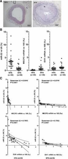
          Expression of miR-22 was closely regulated during VSMC phenotypic modulation. miR-22 overexpression significantly increased expression of VSMC marker genes and inhibited VSMC proliferation and migration, whereas the opposite effect was observed when endogenous miR-22 was knocked down. As expected, 2 previously reported miR-22 target genes, MECP2 (methyl-CpG binding protein 2) and histone deacetylase 4, exhibited a regulatory role in VSMC phenotypic modulation. A transcriptional regulator and oncoprotein, EVI1 (ecotropic virus integration site 1 protein homolog), has been identified as a novel miR-22 target gene in VSMC phenotypic modulation. It is noteworthy that overexpression of miR-22 in the injured vessels significantly reduced the expression of its target genes, decreased VSMC proliferation, and inhibited neointima formation in wire-injured femoral arteries, whereas the opposite effect was observed with local application of a miR-22 inhibitor to injured arteries. We next examined the clinical relevance of miR-22 expression and its target genes in human femoral arteries. We found that miR-22 expression was significantly reduced, whereas MECP2 and EVI1 expression levels were dramatically increased, in diseased in comparison with healthy femoral human arteries. This inverse relationship between miR-22 and MECP2 and EVI1 was evident in both healthy and diseased human femoral arteries.

          Conclusions:

          Our data demonstrate that miR-22 and EVI1 are novel regulators of VSMC function, specifically during neointima hyperplasia, offering a novel therapeutic opportunity for treating vascular diseases.

          Related collections

          Most cited references51

          • Record: found
          • Abstract: found
          • Article: not found

          miR-145 and miR-143 Regulate Smooth Muscle Cell Fate Decisions

          SUMMARY microRNAs are regulators of myriad cellular events, but evidence for a single microRNA that can efficiently differentiate multipotent cells into a specific lineage or regulate direct reprogramming of cells into an alternate cell fate has been elusive. Here, we show that miR-145 and miR-143 are co-transcribed in multipotent cardiac progenitors before becoming localized to smooth muscle cells, including neural crest stem cell–derived vascular smooth muscle cells. miR-145 and miR-143 were direct transcriptional targets of serum response factor, myocardin and Nkx2.5, and were downregulated in injured or atherosclerotic vessels containing proliferating, less differentiated smooth muscle cells. miR-145 was necessary for myocardin-induced reprogramming of adult fibroblasts into smooth muscle cells and sufficient to induce differentiation of multipotent neural crest stem cells into vascular smooth muscle. Furthermore, miR-145 and miR-143 cooperatively targeted a network of transcription factors, including Klf4, myocardin, and Elk-1 to promote differentiation and repress proliferation of smooth muscle cells. These findings demonstrate that miR-145 can direct the smooth muscle fate and that miR-145 and miR-143 function to regulate the quiescent versus proliferative phenotype of smooth muscle cells.
            Bookmark
            • Record: found
            • Abstract: found
            • Article: not found

            Smooth muscle cell phenotypic switching in atherosclerosis.

            Smooth muscle cells (SMCs) possess remarkable phenotypic plasticity that allows rapid adaptation to fluctuating environmental cues, including during development and progression of vascular diseases such as atherosclerosis. Although much is known regarding factors and mechanisms that control SMC phenotypic plasticity in cultured cells, our knowledge of the mechanisms controlling SMC phenotypic switching in vivo is far from complete. Indeed, the lack of definitive SMC lineage-tracing studies in the context of atherosclerosis, and difficulties in identifying phenotypically modulated SMCs within lesions that have down-regulated typical SMC marker genes, and/or activated expression of markers of alternative cell types including macrophages, raise major questions regarding the contributions of SMCs at all stages of atherogenesis. The goal of this review is to rigorously evaluate the current state of our knowledge regarding possible phenotypes exhibited by SMCs within atherosclerotic lesions and the factors and mechanisms that may control these phenotypic transitions.
              Bookmark
              • Record: found
              • Abstract: found
              • Article: not found

              MicroRNA expression signature and antisense-mediated depletion reveal an essential role of MicroRNA in vascular neointimal lesion formation.

              MicroRNAs (miRNAs) are a recently discovered class of endogenous, small, noncoding RNAs that regulate about 30% of the encoding genes of the human genome. However, the role of miRNAs in vascular disease is currently completely unknown. Using microarray analysis, we demonstrated for the first time that miRNAs are aberrantly expressed in the vascular walls after balloon injury. The aberrantly expressed miRNAs were further confirmed by Northern blot and quantitative real-time polymerase chain reaction. Modulating an aberrantly overexpressed miRNA, miR-21, via antisense-mediated depletion (knock-down) had a significant negative effect on neointimal lesion formation. In vitro, the expression level of miR-21 in dedifferentiated vascular smooth muscle cells was significantly higher than that in fresh isolated differentiated cells. Depletion of miR-21 resulted in decreased cell proliferation and increased cell apoptosis in a dose-dependent manner. MiR-21-mediated cellular effects were further confirmed in vivo in balloon-injured rat carotid arteries. Western blot analysis demonstrated that PTEN and Bcl-2 were involved in miR-21-mediated cellular effects. The results suggest that miRNAs are novel regulatory RNAs for neointimal lesion formation. MiRNAs may be a new therapeutic target for proliferative vascular diseases such as atherosclerosis, postangioplasty restenosis, transplantation arteriopathy, and stroke.
                Bookmark

                Author and article information

                Journal
                Circulation
                Circulation
                CIR
                Circulation
                Lippincott Williams & Wilkins
                0009-7322
                1524-4539
                24 April 2018
                23 April 2018
                : 137
                : 17
                : 1824-1841
                Affiliations
                [1 ] 1Department of Cardiology, The First Affiliated Hospital, School of Medicine, Zhejiang University, Hangzhou, China (F.Y., Q.C., M.Y., L.Z.).
                [2 ] 2Centre for Clinical Pharmacology, William Harvey Research Institute, Barts and The London School of Medicine and Dentistry, Queen Mary University of London, United Kingdom (F.Y., S.H., E.M.M., W.A., T.A.A., L.A.L., Q.X.).
                [3 ] 3Key Laboratory of Cardiovascular Diseases, The Second Affiliated Hospital, School of Basic Medical Sciences, Guangzhou Medical University, Xinzao Town, Panyu District, China (Q.X.).
                [4 ] 4Key Laboratory of Protein Modification and Degradation, School of Basic Medical Sciences, Guangzhou Medical University, Xinzao Town, Panyu District, China (Q.X.).
                Author notes
                Li Zhang, MD, PhD, Department of Cardiology, the First Affiliated Hospital, School of Medicine, Zhejiang University, 79 Qingchun Rd, Hangzhou, 310003, Zhejiang, China; or Qingzhong Xiao, MD, PhD, Centre for Clinical Pharmacology, William Harvey Research Institute, Barts and The London School of Medicine and Dentistry, Queen Mary University of London, Heart Centre, Charterhouse Square, London EC1M 6BQ, United Kingdom. E-mail li_zhang@ 123456zju.edu.cn or q.xiao@ 123456qmul.ac.uk
                Article
                00009
                10.1161/CIRCULATIONAHA.117.027799
                5916488
                29246895
                b594275b-5269-41c9-88c0-497e7a336c19
                © 2017 The Authors.

                Circulation is published on behalf of the American Heart Association, Inc., by Wolters Kluwer Health, Inc. This is an open access article under the terms of the Creative Commons Attribution License, which permits use, distribution, and reproduction in any medium, provided that the original work is properly cited.

                History
                : 8 February 2017
                : 4 December 2017
                Categories
                10188
                10192
                10195
                Original Research Articles
                Custom metadata
                TRUE

                atherosclerosis,cell movement,cell proliferation,mds1 and evi1 complex locus protein,methyl-cpg-binding protein 2,micrornas,neointima

                Comments

                Comment on this article