11
views
0
recommends
+1 Recommend
1 collections
    0
    shares
      • Record: found
      • Abstract: found
      • Article: not found

      Recombinant human ACE2: potential therapeutics of SARS-CoV-2 infection and its complication

      research-article
      1 , 2 , 2 , , 1 , 3 ,
      Acta Pharmacologica Sinica
      Springer Singapore

      Read this article at

      ScienceOpenPublisherPMC
      Bookmark
          There is no author summary for this article yet. Authors can add summaries to their articles on ScienceOpen to make them more accessible to a non-specialist audience.

          Abstract

          Dear Editor, Since December 2019, severe acute respiratory syndrome coronavirus 2 (SARS-CoV-2) has caused a pneumonia outbreak in Wuhan city, China, followed by global spread [1, 2]. As of 9 April, 2020, millions of confirmed cases of SARS-CoV-2 infection have been reported, and the global death toll of SARS-CoV-2 infection has surged to tens of thousands of victims, making it a public health emergency of international concern (PHEIC). However, no specific antiviral drug or vaccine for SARS-CoV-2 treatment exists. The high infectivity and the increasing fatality of SARS-CoV-2 highlight the demand for drug discovery. SARS-CoV-2 is closely related to severe acute respiratory syndrome coronavirus (SARS-CoV) [2]. Full-genome sequencing analysis indicated that SARS-CoV-2 shares a high-sequence identity with SARS-CoV [3]. The spike protein (S-protein) of coronaviruses interacts with cell receptors to mediate viral entry into target cells [4]. Additional evidence suggests that both SARS-CoV and SARS-CoV-2 employ angiotensin-converting enzyme 2 (ACE2) as the entry receptor and that the receptor-binding domain (RBD) of the S-protein directly binds to ACE2, triggering endocytosis of virus particles [5–7]. A recent study suggested that the binding affinity between ACE2 and the RBD of SARS-CoV-2 is 10–20 times stronger than that with the RBD of SARS-CoV [5], which likely explains the increased infectivity of SARS-CoV-2. ACE2 is not only a functional receptor of coronaviruses, but also acts as an important negative regulator of the renin–angiotensin system (RAS) through conversion of the vasoconstrictor angiotensin II (Ang II) to its metabolite angiotensin-(1–7) (Ang 1–7) and angiotensin I(Ang I) to angiotensin-(1–9) (Ang 1–9) [7–9]. The ACE2/Ang 1–7 axis plays a series of roles in the improvement of endothelial dysfunction, anti-inflammation, anti-hypertension, anti-thrombus, and anti-fibrosis activity, and cardiovascular protection [10–14]. The protective effect of ACE2 is associated with attenuating Ang II levels and increasing Ang 1–7 levels in lung pathophysiology [10]. Emerging evidence has shown that RAS signaling and ACE2 play crucial roles in SARS-CoV-induced acute respiratory distress syndrome (ARDS) and lethal avian influenza A(H5N1, H7N9)-induced acute lung injury (ALI) [14, 15]. According to pathological findings, SARS-CoV-2 is also associated with lung failure and ARDS [16], and the majority of severely ill patients with SARS-CoV-2 infection have underlying comorbidities, such as cardiovascular disease, diabetes, and cerebrovascular disease [1]. The anti-trypanosomal agent diminazene aceturate (DIZE) was reported to be an ACE2 activator, which has a structure similar to that of the established ACE2 activator xanthenone [17, 18]. DIZE was suggested to exert protective effects in cardiovascular disease through modulating ACE2 activation and expression to increase Ang 1–7 production and improve vascular function [17]. Owing to the role of ACE2 in the entry of SARS-CoV-2, the upregulated expression of ACE2 had an unwanted effect. Therefore, DIZE is not suggested to be applied in the treatment of SARS-CoV-2 infection. However, the addition of exogenous ACE2 could be a potential treatment for SARS-CoV-2 infection, which might not only restrain the spread of SARS-CoV-2 by blocking its interaction with ACE2 on the host cell, but also modulate RAS to treat SARS-CoV-2-related underlying comorbidities and protect the lung from developing ARDS. Given that ACE2 is generated mainly in Clara cells and type II alveolar epithelial cells, the production of ACE2 is severely impaired after epithelial injury in the development of ARDS [19]. In addition, the expression of ACE2 is also severely decreased in patients with pulmonary fibrosis [20]. Therefore, injection of recombinant human ACE2 (rhACE2) is currently considered for treating ARDS and pulmonary arterial hypertension 
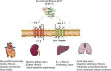
[21]. Circulatory levels of ACE2 activity were markedly increased by rhACE2, which further effectively lowered Ang II levels and generated Ang 1–7 from Ang II (Fig. 1). Although Ang II receptor and ACE blockage were also effective in lung failure in animal models, this treatment could cause potential adverse effects, causing systemic hypotension in humans [22]. As shown in Fig. 1, rhACE2 also acts as a potential therapy for hypertension, heart failure, kidney injury, and liver fibrosis [22–24]. Fig. 1 The mechanism and functions of rhACE2. rhACE2 is able to lower Ang II levels and increase Ang 1–7 levels effectively and exert protective effects in the heart, lung, liver, and kidney. Currently, phase I (NCT00886353) and phase II (NCT01597635) clinical studies with a recombinant version of the catalytic ectodomain of human ACE2 (GSK2586881) have been successfully completed, providing safety and efficacy for ARDS treatment [25, 26]. The administration of rhACE2 was well tolerated without clinically significant hemodynamic changes in healthy subjects and patients with ARDS [26]. During the administration period, no antibodies to rhACE2 were detected, and no serious adverse events were reported [25]. The twice-daily doses of GSK2586881 treatment-regulated angiotensin system peptide, leading to a significant reduction in the concentration of Ang II, accompanied by a rapid rise in Ang 1–7 and Ang 1–5 concentrations, and caused a reduction in IL-6 concentration [26]. However, given the small cohort of critically ill patients, infusion of GSK2586881 did not contribute to ameliorated ARDS through physiological or clinical measures, and a clear role of GSK2586881 in the increased reports of adverse events referring to hypernatremia, pneumonia, dysphagia, and rash was difficult to establish. Therefore, to assess clinical outcomes powerfully, further clinical trials need a larger sample size. Recently, Monteil et al. [27] reported that hrACE2 could significantly inhibit SARS-CoV-2 infection of Vero-E6 cells, and of human capillary and kidney organoids, providing an evidence that rhACE2 might not only reduce lung injury but also block early entry of SARS-CoV-2 infections in target cells. Further studies are needed to illuminate the effect of hrACE2 in SARS-CoV-2 infections from bench to clinic. To ensure the quality of the data and clinical success of rhACE2, the trials for using rhACE2 in patients with SARS-CoV-2 infection or ARDS should consider the patient’s stratification and continuous infusion dose. First, various plasma Ang II levels may pose some difficulties in identifying responders. Hence, before GSK2586881 infusion, the Ang II concentrations and the ratio of ACE2/ACE activity of enrolled patients were evaluated for improved risk stratification. ACE gene insertion/deletion (I/D) polymorphisms play an important role in the development of hypertension, nephritis, and cardiovascular diseases in different ethnic populations by influencing ACE and Ang II activities [28, 29]. Identifying the specific population that is most likely to benefit from rhACE2 represents a bright prospect. Second, due to the short half-life of soluble ACE2 in vivo, a continuous infusion of rhACE2 may enhance efficacy. In addition, an excess of rhACE2 is likely to influence the balance of the RAS; therefore, it is important to identify the effective infusion dose to prevent underlying RAS-related adverse events. Recently, it was reported that a chimeric fusion of rhACE2 and IgG2 Fc fragments could improve rhACE2 plasma stability [22]. This rhACE2-Fc fusion protein retained full peptidase activity and had extended plasma half-life in mice [24]. The strategy for rhACE2-Fc will be expected to provide patients with added convenience, largely reducing administration frequency and greatly improving treatment effectiveness [30]. Taken together, these findings indicate that rhACE2 would represent a potential therapeutic strategy for SARS-CoV-2 infection and its complications.

          Related collections

          Most cited references18

          • Record: found
          • Abstract: found
          • Article: not found

          A Novel Coronavirus from Patients with Pneumonia in China, 2019

          Summary In December 2019, a cluster of patients with pneumonia of unknown cause was linked to a seafood wholesale market in Wuhan, China. A previously unknown betacoronavirus was discovered through the use of unbiased sequencing in samples from patients with pneumonia. Human airway epithelial cells were used to isolate a novel coronavirus, named 2019-nCoV, which formed a clade within the subgenus sarbecovirus, Orthocoronavirinae subfamily. Different from both MERS-CoV and SARS-CoV, 2019-nCoV is the seventh member of the family of coronaviruses that infect humans. Enhanced surveillance and further investigation are ongoing. (Funded by the National Key Research and Development Program of China and the National Major Project for Control and Prevention of Infectious Disease in China.)
            Bookmark
            • Record: found
            • Abstract: found
            • Article: found
            Is Open Access

            A pneumonia outbreak associated with a new coronavirus of probable bat origin

            Since the outbreak of severe acute respiratory syndrome (SARS) 18 years ago, a large number of SARS-related coronaviruses (SARSr-CoVs) have been discovered in their natural reservoir host, bats 1–4 . Previous studies have shown that some bat SARSr-CoVs have the potential to infect humans 5–7 . Here we report the identification and characterization of a new coronavirus (2019-nCoV), which caused an epidemic of acute respiratory syndrome in humans in Wuhan, China. The epidemic, which started on 12 December 2019, had caused 2,794 laboratory-confirmed infections including 80 deaths by 26 January 2020. Full-length genome sequences were obtained from five patients at an early stage of the outbreak. The sequences are almost identical and share 79.6% sequence identity to SARS-CoV. Furthermore, we show that 2019-nCoV is 96% identical at the whole-genome level to a bat coronavirus. Pairwise protein sequence analysis of seven conserved non-structural proteins domains show that this virus belongs to the species of SARSr-CoV. In addition, 2019-nCoV virus isolated from the bronchoalveolar lavage fluid of a critically ill patient could be neutralized by sera from several patients. Notably, we confirmed that 2019-nCoV uses the same cell entry receptor—angiotensin converting enzyme II (ACE2)—as SARS-CoV.
              Bookmark
              • Record: found
              • Abstract: found
              • Article: not found

              Pathological findings of COVID-19 associated with acute respiratory distress syndrome

              Since late December, 2019, an outbreak of a novel coronavirus disease (COVID-19; previously known as 2019-nCoV)1, 2 was reported in Wuhan, China, 2 which has subsequently affected 26 countries worldwide. In general, COVID-19 is an acute resolved disease but it can also be deadly, with a 2% case fatality rate. Severe disease onset might result in death due to massive alveolar damage and progressive respiratory failure.2, 3 As of Feb 15, about 66 580 cases have been confirmed and over 1524 deaths. However, no pathology has been reported due to barely accessible autopsy or biopsy.2, 3 Here, we investigated the pathological characteristics of a patient who died from severe infection with severe acute respiratory syndrome coronavirus 2 (SARS-CoV-2) by postmortem biopsies. This study is in accordance with regulations issued by the National Health Commission of China and the Helsinki Declaration. Our findings will facilitate understanding of the pathogenesis of COVID-19 and improve clinical strategies against the disease. A 50-year-old man was admitted to a fever clinic on Jan 21, 2020, with symptoms of fever, chills, cough, fatigue and shortness of breath. He reported a travel history to Wuhan Jan 8–12, and that he had initial symptoms of mild chills and dry cough on Jan 14 (day 1 of illness) but did not see a doctor and kept working until Jan 21 (figure 1 ). Chest x-ray showed multiple patchy shadows in both lungs (appendix p 2), and a throat swab sample was taken. On Jan 22 (day 9 of illness), the Beijing Centers for Disease Control (CDC) confirmed by reverse real-time PCR assay that the patient had COVID-19. Figure 1 Timeline of disease course according to days from initial presentation of illness and days from hospital admission, from Jan 8–27, 2020 SARS-CoV-2=severe acute respiratory syndrome coronavirus 2. He was immediately admitted to the isolation ward and received supplemental oxygen through a face mask. He was given interferon alfa-2b (5 million units twice daily, atomisation inhalation) and lopinavir plus ritonavir (500 mg twice daily, orally) as antiviral therapy, and moxifloxacin (0·4 g once daily, intravenously) to prevent secondary infection. Given the serious shortness of breath and hypoxaemia, methylprednisolone (80 mg twice daily, intravenously) was administered to attenuate lung inflammation. Laboratory tests results are listed in the appendix (p 4). After receiving medication, his body temperature reduced from 39·0 to 36·4 °C. However, his cough, dyspnoea, and fatigue did not improve. On day 12 of illness, after initial presentation, chest x-ray showed progressive infiltrate and diffuse gridding shadow in both lungs. He refused ventilator support in the intensive care unit repeatedly because he suffered from claustrophobia; therefore, he received high-flow nasal cannula (HFNC) oxygen therapy (60% concentration, flow rate 40 L/min). On day 13 of illness, the patient's symptoms had still not improved, but oxygen saturation remained above 95%. In the afternoon of day 14 of illness, his hypoxaemia and shortness of breath worsened. Despite receiving HFNC oxygen therapy (100% concentration, flow rate 40 L/min), oxygen saturation values decreased to 60%, and the patient had sudden cardiac arrest. He was immediately given invasive ventilation, chest compression, and adrenaline injection. Unfortunately, the rescue was not successful, and he died at 18:31 (Beijing time). Biopsy samples were taken from lung, liver, and heart tissue of the patient. Histological examination showed bilateral diffuse alveolar damage with cellular fibromyxoid exudates (figure 2A, B ). The right lung showed evident desquamation of pneumocytes and hyaline membrane formation, indicating acute respiratory distress syndrome (ARDS; figure 2A). The left lung tissue displayed pulmonary oedema with hyaline membrane formation, suggestive of early-phase ARDS (figure 2B). Interstitial mononuclear inflammatory infiltrates, dominated by lymphocytes, were seen in both lungs. Multinucleated syncytial cells with atypical enlarged pneumocytes characterised by large nuclei, amphophilic granular cytoplasm, and prominent nucleoli were identified in the intra-alveolar spaces, showing viral cytopathic-like changes. No obvious intranuclear or intracytoplasmic viral inclusions were identified. Figure 2 Pathological manifestations of right (A) and left (B) lung tissue, liver tissue (C), and heart tissue (D) in a patient with severe pneumonia caused by SARS-CoV-2 SARS-CoV-2=severe acute respiratory syndrome coronavirus 2. The pathological features of COVID-19 greatly resemble those seen in SARS and Middle Eastern respiratory syndrome (MERS) coronavirus infection.4, 5 In addition, the liver biopsy specimens of the patient with COVID-19 showed moderate microvesicular steatosis and mild lobular and portal activity (figure 2C), indicating the injury could have been caused by either SARS-CoV-2 infection or drug-induced liver injury. There were a few interstitial mononuclear inflammatory infiltrates, but no other substantial damage in the heart tissue (figure 2D). Peripheral blood was prepared for flow cytometric analysis. We found that the counts of peripheral CD4 and CD8 T cells were substantially reduced, while their status was hyperactivated, as evidenced by the high proportions of HLA-DR (CD4 3·47%) and CD38 (CD8 39·4%) double-positive fractions (appendix p 3). Moreover, there was an increased concentration of highly proinflammatory CCR6+ Th17 in CD4 T cells (appendix p 3). Additionally, CD8 T cells were found to harbour high concentrations of cytotoxic granules, in which 31·6% cells were perforin positive, 64·2% cells were granulysin positive, and 30·5% cells were granulysin and perforin double-positive (appendix p 3). Our results imply that overactivation of T cells, manifested by increase of Th17 and high cytotoxicity of CD8 T cells, accounts for, in part, the severe immune injury in this patient. X-ray images showed rapid progression of pneumonia and some differences between the left and right lung. In addition, the liver tissue showed moderate microvesicular steatosis and mild lobular activity, but there was no conclusive evidence to support SARS-CoV-2 infection or drug-induced liver injury as the cause. There were no obvious histological changes seen in heart tissue, suggesting that SARS-CoV-2 infection might not directly impair the heart. Although corticosteroid treatment is not routinely recommended to be used for SARS-CoV-2 pneumonia, 1 according to our pathological findings of pulmonary oedema and hyaline membrane formation, timely and appropriate use of corticosteroids together with ventilator support should be considered for the severe patients to prevent ARDS development. Lymphopenia is a common feature in the patients with COVID-19 and might be a critical factor associated with disease severity and mortality. 3 Our clinical and pathological findings in this severe case of COVID-19 can not only help to identify a cause of death, but also provide new insights into the pathogenesis of SARS-CoV-2-related pneumonia, which might help physicians to formulate a timely therapeutic strategy for similar severe patients and reduce mortality. This online publication has been corrected. The corrected version first appeared at thelancet.com/respiratory on February 25, 2020
                Bookmark

                Author and article information

                Contributors
                cui.pharm@pkufh.com
                yzzhu@must.edu.mo
                Journal
                Acta Pharmacol Sin
                Acta Pharmacol. Sin
                Acta Pharmacologica Sinica
                Springer Singapore (Singapore )
                1671-4083
                1745-7254
                24 June 2020
                : 1-3
                Affiliations
                [1 ]School of Pharmacy and State Key Laboratory for the Quality Research of Chinese Medicine, Macau University of Science and Technology, Macau, China
                [2 ]ISNI 0000 0004 1764 1621, GRID grid.411472.5, Department of Pharmacy, , Peking University First Hospital, ; Beijing, 100034 China
                [3 ]ISNI 0000 0001 0125 2443, GRID grid.8547.e, Shanghai Key Laboratory of Bioactive Small Molecules, Department of Pharmacology, School of Pharmacy, , Fudan University, ; Shanghai, 201203 China
                Article
                430
                10.1038/s41401-020-0430-6
                7313652
                32581256
                b575678b-ee7b-4afe-a4da-22b4112c06bc
                © CPS and SIMM 2020

                This article is made available via the PMC Open Access Subset for unrestricted research re-use and secondary analysis in any form or by any means with acknowledgement of the original source. These permissions are granted for the duration of the World Health Organization (WHO) declaration of COVID-19 as a global pandemic.

                History
                : 5 March 2020
                : 22 April 2020
                Categories
                Correspondence

                Pharmacology & Pharmaceutical medicine
                Pharmacology & Pharmaceutical medicine

                Comments

                Comment on this article