23
views
0
recommends
+1 Recommend
0 collections
    0
    shares
      • Record: found
      • Abstract: found
      • Article: found
      Is Open Access

      High Affinity Neurexin Binding to Cell Adhesion G-protein-coupled Receptor CIRL1/Latrophilin-1 Produces an Intercellular Adhesion Complex*

      research-article

      Read this article at

      Bookmark
          There is no author summary for this article yet. Authors can add summaries to their articles on ScienceOpen to make them more accessible to a non-specialist audience.

          Abstract

          Background: Neurexins and CIRL/latrophilin-1 (CL1) are independent synaptic receptors for α-latrotoxin.

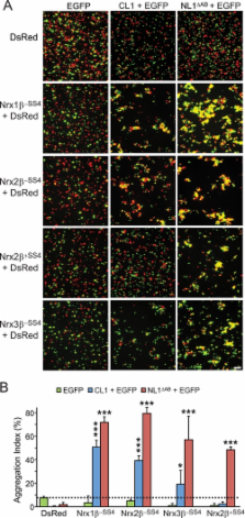
          Results: Neurexins and CL1 form a high affinity complex that mediates intercellular adhesion and is regulated by neurexin alternative splicing.

          Conclusion: Thus, two independent α-latrotoxin receptors interact trans-cellularly to form a connection between neurons.

          Significance: The neurexin-CL1 complex may be involved in trans-synaptic cell adhesion and mediate α-latrotoxin toxicity.

          Abstract

          The G-protein-coupled receptor CIRL1/latrophilin-1 (CL1) and the type-1 membrane proteins neurexins represent distinct neuronal cell adhesion molecules that exhibit no similarities except for one common function: both proteins are receptors for α-latrotoxin, a component of black widow spider venom that induces massive neurotransmitter release at synapses. Unexpectedly, we have now identified a direct binding interaction between the extracellular domains of CL1 and neurexins that is regulated by alternative splicing of neurexins at splice site 4 (SS4). Using saturation binding assays, we showed that neurexins lacking an insert at SS4 bind to CL1 with nanomolar affinity, whereas neurexins containing an insert at SS4 are unable to bind. CL1 competed for neurexin binding with neuroligin-1, a well characterized neurexin ligand. The extracellular sequences of CL1 contain five domains (lectin, olfactomedin-like, serine/threonine-rich, hormone-binding, and G-protein-coupled receptor autoproteolysis-inducing (GAIN) domains). Of these domains, the olfactomedin-like domain mediates neurexin binding as shown by deletion mapping. Cell adhesion assays using cells expressing neurexins and CL1 revealed that their interaction produces a stable intercellular adhesion complex, indicating that their interaction can be trans-cellular. Thus, our data suggest that CL1 constitutes a novel ligand for neurexins that may be localized postsynaptically based on its well characterized interaction with intracellular SH3 and multiple ankyrin repeats adaptor proteins (SHANK) and could form a trans-synaptic complex with presynaptic neurexins.

          Related collections

          Most cited references48

          • Record: found
          • Abstract: found
          • Article: not found

          Neuroligin expressed in nonneuronal cells triggers presynaptic development in contacting axons.

          Most neurons form synapses exclusively with other neurons, but little is known about the molecular mechanisms mediating synaptogenesis in the central nervous system. Using an in vitro system, we demonstrate that neuroligin-1 and -2, postsynaptically localized proteins, can trigger the de novo formation of presynaptic structure. Nonneuronal cells engineered to express neuroligins induce morphological and functional presynaptic differentiation in contacting axons. This activity can be inhibited by addition of a soluble version of beta-neurexin, a receptor for neuroligin. Furthermore, addition of soluble beta-neurexin to a coculture of defined pre- and postsynaptic CNS neurons inhibits synaptic vesicle clustering in axons contacting target neurons. Our results suggest that neuroligins are part of the machinery employed during the formation and remodeling of CNS synapses.
            Bookmark
            • Record: found
            • Abstract: found
            • Article: not found

            Neurexins induce differentiation of GABA and glutamate postsynaptic specializations via neuroligins.

            Formation of synaptic connections requires alignment of neurotransmitter receptors on postsynaptic dendrites opposite matching transmitter release sites on presynaptic axons. beta-neurexins and neuroligins form a trans-synaptic link at glutamate synapses. We show here that neurexin alone is sufficient to induce glutamate postsynaptic differentiation in contacting dendrites. Surprisingly, neurexin also induces GABA postsynaptic differentiation. Conversely, neuroligins induce presynaptic differentiation in both glutamate and GABA axons. Whereas neuroligins-1, -3, and -4 localize to glutamate postsynaptic sites, neuroligin-2 localizes primarily to GABA synapses. Direct aggregation of neuroligins reveals a linkage of neuroligin-2 to GABA and glutamate postsynaptic proteins, but the other neuroligins only to glutamate postsynaptic proteins. Furthermore, mislocalized expression of neuroligin-2 disperses postsynaptic proteins and disrupts synaptic transmission. Our findings indicate that the neurexin-neuroligin link is a core component mediating both GABAergic and glutamatergic synaptogenesis, and differences in isoform localization and binding affinities may contribute to appropriate differentiation and specificity.
              Bookmark
              • Record: found
              • Abstract: found
              • Article: not found

              Alpha-neurexins couple Ca2+ channels to synaptic vesicle exocytosis.

              Synapses are specialized intercellular junctions in which cell adhesion molecules connect the presynaptic machinery for neurotransmitter release to the postsynaptic machinery for receptor signalling. Neurotransmitter release requires the presynaptic co-assembly of Ca2+ channels with the secretory apparatus, but little is known about how synaptic components are organized. Alpha-neurexins, a family of >1,000 presynaptic cell-surface proteins encoded by three genes, link the pre- and postsynaptic compartments of synapses by binding extracellularly to postsynaptic cell adhesion molecules and intracellularly to presynaptic PDZ domain proteins. Using triple-knockout mice, we show that alpha-neurexins are not required for synapse formation, but are essential for Ca2+-triggered neurotransmitter release. Neurotransmitter release is impaired because synaptic Ca2+ channel function is markedly reduced, although the number of cell-surface Ca2+ channels appears normal. These data suggest that alpha-neurexins organize presynaptic terminals by functionally coupling Ca2+ channels to the presynaptic machinery.
                Bookmark

                Author and article information

                Journal
                J Biol Chem
                J. Biol. Chem
                jbc
                jbc
                JBC
                The Journal of Biological Chemistry
                American Society for Biochemistry and Molecular Biology (9650 Rockville Pike, Bethesda, MD 20814, U.S.A. )
                0021-9258
                1083-351X
                16 March 2012
                19 January 2012
                19 January 2012
                : 287
                : 12
                : 9399-9413
                Affiliations
                [1]From the Department of Molecular and Cellular Physiology and Howard Hughes Medical Institute, Stanford University School of Medicine, Stanford, California 94305
                Author notes
                [1 ] To whom correspondence may be addressed: Dept. of Molecular and Cellular Physiology and Howard Hughes Medical Inst., Stanford University School of Medicine, 265 Campus Dr., Stanford, CA 94305. E-mail: aboucard@ 123456stanford.edu .
                [3 ] To whom correspondence may be addressed: Dept. of Molecular and Cellular Physiology and Howard Hughes Medical Inst., Stanford University School of Medicine, 265 Campus Dr., Stanford, CA 94305. E-mail: tcs1@ 123456stanford.edu .
                [2]

                Present address: Dept. of Biochemistry, College of Life Science and Biotechnology, Yonsei University, 134 Shinchon-dong, Seodaemun-gu, Science Research Center S421, Seoul 120-749, South Korea.

                Article
                M111.318659
                10.1074/jbc.M111.318659
                3308797
                22262843
                b37eafd6-c005-4df8-a025-fd22f4189a79
                © 2012 by The American Society for Biochemistry and Molecular Biology, Inc.

                Author's Choice—Final version full access.

                Creative Commons Attribution Non-Commercial License applies to Author Choice Articles

                History
                : 28 October 2011
                : 9 January 2012
                Funding
                Funded by: National Institutes of Health
                Award ID: R37 MH052804-18
                Categories
                Neurobiology

                Biochemistry
                neurotoxin,receptor structure-function,cell adhesion,cell surface receptor,g-protein-coupled receptors (gpcr),synapses,autism,protein/protein interactions,neuroscience,neurological diseases

                Comments

                Comment on this article