7
views
0
recommends
+1 Recommend
0 collections
    0
    shares
      • Record: found
      • Abstract: found
      • Article: found
      Is Open Access

      Characterization and noninvasive diagnosis of bladder cancer with serum surface enhanced Raman spectroscopy and genetic algorithms

      research-article

      Read this article at

      Bookmark
          There is no author summary for this article yet. Authors can add summaries to their articles on ScienceOpen to make them more accessible to a non-specialist audience.

          Abstract

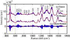
          This study aims to characterize and classify serum surface-enhanced Raman spectroscopy (SERS) spectra between bladder cancer patients and normal volunteers by genetic algorithms (GAs) combined with linear discriminate analysis (LDA). Two group serum SERS spectra excited with nanoparticles are collected from healthy volunteers (n = 36) and bladder cancer patients (n = 55). Six diagnostic Raman bands in the regions of 481–486, 682–687, 1018–1034, 1313–1323, 1450–1459 and 1582–1587 cm −1 related to proteins, nucleic acids and lipids are picked out with the GAs and LDA. By the diagnostic models built with the identified six Raman bands, the improved diagnostic sensitivity of 90.9% and specificity of 100% were acquired for classifying bladder cancer patients from normal serum SERS spectra. The results are superior to the sensitivity of 74.6% and specificity of 97.2% obtained with principal component analysis by the same serum SERS spectra dataset. Receiver operating characteristic (ROC) curves further confirmed the efficiency of diagnostic algorithm based on GA-LDA technique. This exploratory work demonstrates that the serum SERS associated with GA-LDA technique has enormous potential to characterize and non-invasively detect bladder cancer through peripheral blood.

          Related collections

          Most cited references22

          • Record: found
          • Abstract: found
          • Article: not found

          Surface-enhanced Raman spectroscopy (SERS): progress and trends.

          Surface-enhanced Raman spectroscopy (SERS) combines molecular fingerprint specificity with potential single-molecule sensitivity. Therefore, the SERS technique is an attractive tool for sensing molecules in trace amounts within the field of chemical and biochemical analytics. Since SERS is an ongoing topic, which can be illustrated by the increased annual number of publications within the last few years, this review reflects the progress and trends in SERS research in approximately the last three years. The main reason why the SERS technique has not been established as a routine analytic technique, despite its high specificity and sensitivity, is due to the low reproducibility of the SERS signal. Thus, this review is dominated by the discussion of the various concepts for generating powerful, reproducible, SERS-active surfaces. Furthermore, the limit of sensitivity in SERS is introduced in the context of single-molecule spectroscopy and the calculation of the 'real' enhancement factor. In order to shed more light onto the underlying molecular processes of SERS, the theoretical description of SERS spectra is also a growing research field and will be summarized here. In addition, the recording of SERS spectra is affected by a number of parameters, such as laser power, integration time, and analyte concentration. To benefit from synergies, SERS is combined with other methods, such as scanning probe microscopy and microfluidics, which illustrates the broad applications of this powerful technique.
            Bookmark
            • Record: found
            • Abstract: found
            • Article: not found

            Real-time Raman spectroscopy for in vivo skin cancer diagnosis.

            Raman spectroscopy is a noninvasive optical technique capable of measuring vibrational modes of biomolecules within viable tissues. In this study, we evaluated the application of an integrated real-time system of Raman spectroscopy for in vivo skin cancer diagnosis. Benign and malignant skin lesions (n = 518) from 453 patients were measured within 1 second each, including melanomas, basal cell carcinomas, squamous cell carcinomas, actinic keratoses, atypical nevi, melanocytic nevi, blue nevi, and seborrheic keratoses. Lesion classification was made using a principal component with general discriminant analysis and partial least-squares in three distinct discrimination tasks: skin cancers and precancers from benign skin lesions [receiver operating characteristic (ROC) = 0.879]; melanomas from nonmelanoma pigmented lesions (ROC = 0.823); and melanomas from seborrheic keratoses (ROC = 0.898). For sensitivities between 95% and 99%, the specificities ranged between 15% and 54%. Our findings establish that real-time Raman spectroscopy can be used to distinguish malignant from benign skin lesions with good diagnostic accuracy comparable with clinical examination and other optical-based methods. ©2012 AACR.
              Bookmark
              • Record: found
              • Abstract: found
              • Article: not found

              Nasopharyngeal cancer detection based on blood plasma surface-enhanced Raman spectroscopy and multivariate analysis.

              A surface-enhanced Raman spectroscopy (SERS) method was developed for blood plasma biochemical analysis for the first time with the aim to develop a simple blood test for non-invasive nasopharyngeal cancer detection. Silver nanoparticles (Ag NP) as the SERS-active nanostructures were directly mixed with blood plasma to enhance the Raman scattering signals of various biomolecular constituents such as proteins, lipids, and nucleic acids. High quality SERS spectrum from blood plasma-Ag NP mixture can be obtained within 10s using a Renishaw micro-Raman system. SERS measurements were performed on two groups of blood plasma samples: one group from patients (n=43) with pathologically confirmed nasopharyngeal carcinomas (WHO type I, II, and III) and the other group from healthy volunteers (control subjects, n=33). Tentative assignments of the Raman bands in the measured SERS spectra suggest interesting cancer specific biomolecular differences, including an increase in the relative amounts of nucleic acid, collagen, phospholipids and phenylalanine and a decrease in the percentage of amino acids and saccharide contents in the blood plasma of nasopharyngeal cancer patients as compared to that of healthy subjects. Principal component analysis (PCA) of the measured SERS spectra separated the spectral features of the two groups into two distinct clusters with little overlaps. Linear discriminate analysis (LDA) based on the PCA generated features differentiated the nasopharyngeal cancer SERS spectra from normal SERS spectra with high sensitivity (90.7%) and specificity (100%). The results from this exploratory study demonstrated great potentials for developing SERS blood plasma analysis into a novel clinical tool for non-invasive detection of nasopharyngeal cancers.
                Bookmark

                Author and article information

                Journal
                Sci Rep
                Sci Rep
                Scientific Reports
                Nature Publishing Group
                2045-2322
                07 May 2015
                2015
                : 5
                : 9582
                Affiliations
                [1 ]Biomedical Engineering Laboratory, School of Information Engineering, Guangdong Medical College , Dongguan 523808, Guangdong, China
                [2 ]State Key Laboratory of Oncology in South China and Department of Clinical Laboratory, Sun Yat-sen University Cancer Center , Guangzhou, 510060, China
                [3 ]School of Basic Medicine, Guangdong Medical College , Dongguan 523808, Guangdong, China
                [4 ]MOE Key Laboratory of Laser Life Science & SATCM Third Grade Laboratory of Chinese Medicine and Photonics Technology, College of Biophotonics, South China Normal University , Guangzhou 510631, China
                [5 ]Guangdong Provincial Key Laboratory of Medical Molecular Diagnostics , No. 1 Xincheng Road, Dongguan 523808, China
                Author notes
                [*]

                These authors contributed equally to this work.

                Article
                srep09582
                10.1038/srep09582
                4423238
                25947114
                b349f957-335b-44f7-b9c6-44ba4dc3a2c2
                Copyright © 2015, Macmillan Publishers Limited. All rights reserved

                This work is licensed under a Creative Commons Attribution 4.0 International License. The images or other third party material in this article are included in the article's Creative Commons license, unless indicated otherwise in the credit line; if the material is not included under the Creative Commons license, users will need to obtain permission from the license holder in order to reproduce the material. To view a copy of this license, visit http://creativecommons.org/licenses/by/4.0/

                History
                : 12 November 2014
                : 04 March 2015
                Categories
                Article

                Uncategorized
                Uncategorized

                Comments

                Comment on this article