14
views
0
recommends
+1 Recommend
0 collections
    0
    shares
      • Record: found
      • Abstract: found
      • Article: found
      Is Open Access

      Native architecture of the Chlamydomonas chloroplast revealed by in situ cryo-electron tomography

      research-article

      Read this article at

      Bookmark
          There is no author summary for this article yet. Authors can add summaries to their articles on ScienceOpen to make them more accessible to a non-specialist audience.

          Abstract

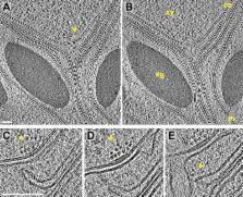
          Chloroplast function is orchestrated by the organelle's intricate architecture. By combining cryo-focused ion beam milling of vitreous Chlamydomonas cells with cryo-electron tomography, we acquired three-dimensional structures of the chloroplast in its native state within the cell. Chloroplast envelope inner membrane invaginations were frequently found in close association with thylakoid tips, and the tips of multiple thylakoid stacks converged at dynamic sites on the chloroplast envelope, implicating lipid transport in thylakoid biogenesis. Subtomogram averaging and nearest neighbor analysis revealed that RuBisCO complexes were hexagonally packed within the pyrenoid, with ∼15 nm between their centers. Thylakoid stacks and the pyrenoid were connected by cylindrical pyrenoid tubules, physically bridging the sites of light-dependent photosynthesis and light-independent carbon fixation. Multiple parallel minitubules were bundled within each pyrenoid tubule, possibly serving as conduits for the targeted one-dimensional diffusion of small molecules such as ATP and sugars between the chloroplast stroma and the pyrenoid matrix.

          DOI: http://dx.doi.org/10.7554/eLife.04889.001

          eLife digest

          Many organisms can harvest light to produce their own energy through a process called photosynthesis. In plant and algal cells, photosynthesis takes place within the chloroplasts, which are compartments that contain stacks of structures called thylakoids.

          Inside the thylakoids, proteins absorb energy from light and convert it into biochemical energy that can be used by the cell. This energy then powers a series of reactions that result in carbon dioxide being incorporated into energy-rich sugars. The enzyme RuBisCO is essential for this process, and is believed to be the most abundant protein on Earth. In land plants, RuBisCO is found throughout the chloroplast, but in algae it is limited to a specialized area called the pyrenoid.

          Much of our current knowledge of chloroplast structure comes from transmission electron microscopy (TEM) images. However, the traditional methods used to prepare cells for TEM can damage their internal structures. Also, previous studies have focused primarily on the chloroplasts of land plants, even though aquatic organisms—including the alga Chlamydomonas—account for over 50% of photosynthesis on the planet.

          Here, Engel et al. provide the first three-dimensional structures of Chlamydomonas chloroplasts in their natural state. They used several recently-developed techniques to study cells that were preserved in a close-to-living condition. The cells were rapidly frozen, thinned with a technique called cryo-focused ion beam milling, and then imaged by a type of TEM called cryo-electron tomography.

          The three-dimensional images provide many insights into the Chlamydomonas chloroplast, including evidence that lipids and proteins move between the membrane that surrounds the chloroplast—called the chloroplast envelope—and the tips of the thylakoids. These images show how thylakoids may be built by the transport of molecules from the chloroplast envelope. In addition, the images reveal the detailed structures of the tubes that connect the thylakoids to the pyrenoid, which could explain how the two stages of photosynthesis (light harvesting and the conversion of carbon dioxide) can be coordinated even though they occur at different places within the chloroplast.

          Engel et al. also observed that RuBisCO enzymes are arranged in a hexagonal pattern inside the pyrenoid, but are spaced too far apart to make direct contact with each other. To understand how the pyrenoid is assembled, a future goal will be to determine what causes RuBisCO to be arranged in this way.

          DOI: http://dx.doi.org/10.7554/eLife.04889.002

          Related collections

          Most cited references104

          • Record: found
          • Abstract: not found
          • Article: not found

          Multidimensional binary search trees used for associative searching

            Bookmark
            • Record: found
            • Abstract: not found
            • Article: not found

            Primary Production of the Biosphere: Integrating Terrestrial and Oceanic Components

            C Field (1998)
              Bookmark
              • Record: found
              • Abstract: found
              • Article: not found

              Focused-ion-beam thinning of frozen-hydrated biological specimens for cryo-electron microscopy.

              Cryo-electron microscopy can provide high-resolution structural information about cells and organelles in the nearly native, frozen-hydrated state. Applicability, however, is limited by difficulties encountered in preparing suitably thin, vitreously frozen biological specimens. We demonstrate, by cryo-electron tomography of Escherichia coli cells, that a focused ion beam (FIB) can be used to thin whole frozen-hydrated cells in a convenient and essentially artifact-free way.
                Bookmark

                Author and article information

                Contributors
                Role: Reviewing editor
                Journal
                eLife
                eLife
                eLife
                eLife
                eLife Sciences Publications, Ltd
                2050-084X
                2050-084X
                13 January 2015
                2015
                : 4
                : e04889
                Affiliations
                [1 ]deptDepartment of Molecular Structural Biology , Max Planck Institute of Biochemistry , Martinsried, Germany
                Salk Institute , United States
                Salk Institute , United States
                Author notes
                [* ]For correspondence: engelben@ 123456biochem.mpg.de (BDE);
                [†]

                These authors contributed equally to this work.

                [‡]

                Department of Chemistry and Biochemistry, University of California, San Diego, La Jolla, United States.

                [§]

                Department of Chemistry, Bijvoet Center for Biomolecular Research, Utrecht University, Utrecht, Netherlands.

                Article
                04889
                10.7554/eLife.04889
                4292175
                25584625
                b260910c-1c9e-46ca-9a6b-97bc278967df
                © 2014, Engel et al

                This article is distributed under the terms of the Creative Commons Attribution License, which permits unrestricted use and redistribution provided that the original author and source are credited.

                History
                : 24 September 2014
                : 08 December 2014
                Funding
                Funded by: FundRef http://dx.doi.org/10.13039/501100000780, European Commission;
                Award ID: 7th Framework Programme Grant HEALTH-F4-2008-201648/PROSPECTS
                Award Recipient :
                Funded by: FundRef http://dx.doi.org/10.13039/100005156, Alexander von Humboldt-Stiftung (Humboldt Foundation);
                Award ID: Postdoctoral Research Fellowship
                Award Recipient :
                Funded by: FundRef http://dx.doi.org/10.13039/501100001655, German Academic Exchange Service;
                Award ID: CONACyT-DAAD Graduate Scholarship
                Award Recipient :
                Funded by: FundRef http://dx.doi.org/10.13039/501100001659, Deutsche Forschungsgemeinschaft;
                Award ID: Excellence Clusters CIPSM and SFB 1035
                Award Recipient :
                Funded by: FundRef http://dx.doi.org/10.13039/501100002347, Bundesministerium für Bildung und Forschung (Federal Ministry of Education and Research);
                Award Recipient :
                Funded by: FundRef http://dx.doi.org/10.13039/501100004189, Max-Planck-Gesellschaft (Max Planck Society);
                Award ID: Inter-Institutional Research Initiative
                Award Recipient :
                The funders had no role in study design, data collection and interpretation, or the decision to submit the work for publication.
                Categories
                Research Article
                Biophysics and Structural Biology
                Plant Biology
                Custom metadata
                2.0
                The first 3D views of the native algal chloroplast provide new insights into thylakoid biogenesis, photosynthesis, and carbon fixation.

                Life sciences
                chlamydomonas,focused ion beam,cryo-electron tomography,chloroplast,thylakoid,rubisco,other
                Life sciences
                chlamydomonas, focused ion beam, cryo-electron tomography, chloroplast, thylakoid, rubisco, other

                Comments

                Comment on this article