92
views
0
recommends
+1 Recommend
0 collections
    0
    shares
      • Record: found
      • Abstract: found
      • Article: found
      Is Open Access

      Laparoscopic diagnosis and treatment of a hydrocele of the canal of Nuck extending in the retroperitoneal space: A case report

      other

      Read this article at

      Bookmark
          There is no author summary for this article yet. Authors can add summaries to their articles on ScienceOpen to make them more accessible to a non-specialist audience.

          Highlights

          • Hydrocele of the canal of Nuck in the adult female is a rare condition.

          • Laparoscopic diagnosis is useful for groin mass connected with the retroperitoneal space.

          • TEP offers a useful alternative in selected patients with hydrocele of the canal of Nuck.

          Abstract

          INTRODUCTION

          Hydrocele of the canal of Nuck is a rarely encountered entity. We report a case underwent laparoscopic totally extraperitoneal (TEP) treatment for a hydrocele of the canal of Nuck extending in the extraperitoneal space mainly.

          PRESENTATION OF CASE

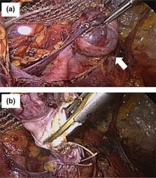
          A 37-year-old woman complained of painless and reducible swelling in her left groin, and referred to our hospital for surgical management against left inguinal hernia with the incarcerated ovary. Ultrasonography and MR images revealed a cystic mass in the retroperitoneal space, and we diagnosed as an unusual type of hydrocele of the canal of Nuck. The patient was scheduled for laparoscopic treatment. Laparoscopic findings on pneumoperitoneum showed an extraperitoneal cystic tumor with no contact with the left ovary. The fascia and peritoneum of the port site were closed, and then an extraperitoneal space was created. The cystic tumor with the round ligament of the uterus was dissected and resected by the TEP technique. The extended deep inguinal ring was repaired with polypropylene mesh. Postoperative course was uneventful.

          DISCUSSION

          Hydrocele of the canal of Nuck in the adult female is a rare condition. The accurate diagnosis of an inguinal hydrocele in a female is seldom made. Laparoscopic examination provides surgeons with information of inguinal swelling accompanied with retroperitoneal cyst, and consecutive treatment by laparoscopic technique, especially TEP, is useful in regard to minimal damage of the peritoneum.

          CONCLUSION

          Laparoscopic diagnosis and TEP treatment offers a useful alternative in selected patients with hydrocele of the canal of Nuck.

          Related collections

          Most cited references13

          • Record: found
          • Abstract: found
          • Article: not found

          Comparison of conventional anterior surgery and laparoscopic surgery for inguinal-hernia repair.

          Inguinal hernias can be repaired by laparoscopic techniques, which have had better results than open surgery in several small studies. We performed a randomized, multicenter trial in which 487 patients with inguinal hernias were treated by extraperitoneal laparoscopic repair and 507 patients were treated by conventional anterior repair. We recorded information about postoperative recovery and complications and examined the patients for recurrences one and six weeks, six months, and one and two years after surgery. Six patients in the open-surgery group but none in the laparoscopic-surgery group had wound abscesses (P=0.03), and the patients in the laparoscopic-surgery group had a more rapid recovery (median time to the resumption of normal daily activity, 6 vs. 10 days; time to the return to work, 14 vs. 21 days; and time to the resumption of athletic activities, 24 vs. 36 days; P<0.001 for all comparisons). With a median follow-up of 607 days, 31 patients (6 percent) in the open-surgery group had recurrences, as compared with 17 patients (3 percent) in the laparoscopic-surgery group (P=0.05). All but three of the recurrences in the latter group were within one year after surgery and were caused by surgeon-related errors. In the open-surgery group, 15 patients had recurrences during the first year, and 16 during the second year. Follow-up was complete for 97 percent of the patients. Patients with inguinal hernias who undergo laparoscopic repair recover more rapidly and have fewer recurrences than those who undergo open surgical repair.
            Bookmark
            • Record: found
            • Abstract: found
            • Article: not found

            Imaging of groin masses: inguinal anatomy and pathologic conditions revisited.

            The groin region is subdivided into two distinct anatomic areas: the inguinal canal and the femoral triangle. A series of cross-sectional imaging cases illustrate that a good understanding of the local anatomic characteristics of the groin allows confident characterization of groin pathologic conditions. Such conditions can be classified into five major groups: congenital abnormalities, noncongenital hernias, vascular conditions, infectious or inflammatory processes, and neoplasms. Congenital entities include hernias, cysts, undescended testis, and retractile testes. Ultrasound (US) is useful in depicting these conditions. In the second group, noncongenital hernias, US allows visualization of bowel loops in peristalsis within the hernia. Herniography, computed tomography (CT), and magnetic resonance (MR) imaging are also helpful in diagnosis. Among vascular conditions, false aneurysms may be detected from the turbulent flow through the tract at Doppler US. The characteristic venous flow of varicoceles is best diagnosed with US during the Valsalva maneuver. Infectious or inflammatory conditions include, among others, hematomas, which appear hyperattenuating at CT and have variable appearances, depending on the age of the blood products, at MR imaging. The origins of neoplasms may be assessed at CT and MR imaging, although appearances of solid tumors are relatively nonspecific.
              Bookmark
              • Record: found
              • Abstract: found
              • Article: not found

              The presentation of asymptomatic palpable movable mass in female inguinal hernia.

              The surgical approach to the inguinal canal in girls is identical to that in boys. The sliding hernia which contains the ovary with or without the fallopian tube occurs occasionally in female patients. In our clinical experience, we found that a hydrocele in the labium, also presenting with an asymptomatic palpable movable mass, mimics a sliding hernia with ovary. In an attempt to differentiate between hydrocele and sliding inguinal hernia with ovary in female patients, we report our experience dealing with the two situations at a single institution within a 5-year period. Between July 1997 and June 2002, 1800 infants and children underwent surgery for inguinal hernia at Chang Gung Children's Hospital, of whom 580 were female infants and girls aged 1 month to 14 years (mean, 5.7 years). Some 32 patients (5.3%) presented with an asymptomatic palpable movable mass over the labium major. Pre-operative sonography was performed for all cases. Twenty-six female infants aged 1 month to 18 months (mean 5 months) had sliding hernia; both the ovary and fallopian tube were contained. Six girls aged 2 years to 6 years (mean 4.6 years), had hydrocele of the canal of Nuck. The accuracy of pre-operative diagnosis with sonography was 100%. Sonography is an easy and accurate pre-operative diagnostic procedure. We suggest that sonography be performed routinely in all female cases with an inguinal hernia containing a palpable movable mass.
                Bookmark

                Author and article information

                Contributors
                Journal
                Int J Surg Case Rep
                Int J Surg Case Rep
                International Journal of Surgery Case Reports
                Elsevier
                2210-2612
                08 October 2014
                08 October 2014
                2014
                : 5
                : 11
                : 861-864
                Affiliations
                [0005]Department of Surgery, National Hospital Organization Beppu Medical Center, Oita, Japan
                Author notes
                [* ]Corresponding author at: Department of Surgery, National Hospital Organization Beppu Medical Center, 1473 Uchikamado, Beppu, Oita 874-0011, Japan. Tel.: +81 977 67 1111; fax: +81 977 67 5766. tmatsu@ 123456beppu.hosp.go.jp
                Article
                S2210-2612(14)00197-7
                10.1016/j.ijscr.2014.08.016
                4245671
                25462052
                b14c0d48-7801-4a31-87a1-1e612157526d
                © 2014 The Authors

                This is an open access article under the CC BY-NC-ND license (http://creativecommons.org/licenses/by-nc-nd/3.0/).

                History
                : 17 April 2014
                : 19 July 2014
                : 16 August 2014
                Categories
                Article

                laparoscopy,the canal of nuck,hydrocele,tep
                laparoscopy, the canal of nuck, hydrocele, tep

                Comments

                Comment on this article