4
views
0
recommends
+1 Recommend
1 collections
    0
    shares
      • Record: found
      • Abstract: found
      • Article: found
      Is Open Access

      BONE TUNNEL ENLARGEMENT WITH NON-METALLIC INTERFERENCE SCREWS IN ACL RECONSTRUCTION Translated title: ALARGAMENTO DOS TÚNEIS ÓSSEOS NA RECONSTRUÇÃO DO LCA COM PARAFUSOS DE INTERFERÊNCIA NÃO METÁLICOS

      research-article

      Read this article at

      ScienceOpenPublisherPMC
      Bookmark
          There is no author summary for this article yet. Authors can add summaries to their articles on ScienceOpen to make them more accessible to a non-specialist audience.

          ABSTRACT

          Objective

          To compare the widening of bone tunnels between poly-etheretherketone (PEEK), absorbable polylactic acid DL (PLDL) and tricalcium phosphate (TCP) interference screws in anterior cruciate ligament (ACL) reconstruction.

          Methods

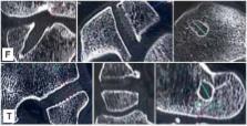
          Three groups of patients undergoing ACL reconstruction with at least 1 year of follow-up using the out-in drilling technique and hamstring as a graft were assessed. The patients were divided according to the type of interference screw used (PEEK, PLDL and TCP). Computed tomography (CT) was performed to measure the greatest femoral and tibial tunnel widening regarding to the initial tunnel, and then it was compared between groups.

          Results

          Mean widening in group 1 (PEEK) was 39.56% (SD 16%) in the femoral tunnel and 33.65% (SD 20%) in the tibia. In group 2 (PLDL) mean widening was 48.43% in the femoral tunnel (SD 18%) and 35.24% (SD 13%) in the tibial tunnel. In group 3 (TCP) mean widening was 44.51% in the femur (SD 14%) and 36.83% in the tibia (SD 14%). The comparison between groups (PLDL-PEEK, PLDL-TCP, PEEK-TCP) shows no statistically significant difference.

          Conclusion

          Bone tunnel enlargement values after ACL reconstruction with the use of different types of materials (bioinert and biomaterials) of interference screws (PEEK, PLDL and TCP) were similar. Level of Evidence III, Comparative retrospective study.

          RESUMO

          Objetivo

          Comparar o alargamento dos túneis ósseos entre parafusos de interferência de poli-éter-etil-cetona (PEEK), ácido poli lático (PLDL) absorvível e tricálcio fosfato (TCP) na reconstrução do ligamento cruzado anterior (LCA).

          Métodos

          Foram avaliados três grupos de pacientes submetidos à reconstrução do LCA com ao menos um ano de acompanhamento, com perfuração de fora para dentro, tendões flexores quádruplos como enxerto, que foram divididos de acordo com o parafuso de interferência utilizado (PEEK, PLDL e TCP). Realizou-se tomografia computadorizada (TC) para aferição do maior alargamento do túnel tibial e femoral em relação ao túnel inicial, e foi comparado o alargamento entre os grupos.

          Resultados

          O alargamento médio no grupo 1 (PEEK) foi 39,56% (DP = 16%) no túnel femoral e 33,65% (DP = 20%) na tíbia. No grupo 2 (PLDL) o alargamento médio do túnel femoral foi 48,43% (DP = 18%) e 35,24% (DP = 13%) na tíbia. No grupo 3 (TCP) 44,51% (DP = 14%) foi o alargamento médio no fêmur e 36.83% (DP = 14%) na tíbia. Na comparação entre os grupos (PLDL-PEEK, PLDL-TCP, PEEK-TCP) não houve diferença estatisticamente significante.

          Conclusão

          O alargamento dos túneis ósseos após a reconstrução do LCA com a utilização de diferentes tipos de materiais (bioinertes e biomateriais) de parafusos de interferência (PEEK, PLDL e TCP) foi semelhante. Nível de Evidencia III, Estudo retrospectivo comparativo.

          Related collections

          Most cited references21

          • Record: found
          • Abstract: found
          • Article: not found

          A prospective evaluation of tunnel enlargement in anterior cruciate ligament reconstruction with hamstrings: extracortical versus anatomical fixation.

          Changes in the femoral and tibial bone tunnel were studied prospectively after arthroscopic ACL reconstruction with quadruple hamstring autograft. To determine whether tunnel enlargement can be decreased by fixing the graft close to the joint line having a stiffer fixation construct we compared "anatomical" (one absorbable interference screw femorally, and bicortical fixation with two absorbable interference screws tibially) and extracortical fixation techniques (Endobutton femorally, and two no. 6 Ethibond sutures over a suture washer tibially). Over a 2-year period we evaluated 60 patients clinically (IKDC scale, Cincinnati Knee Score, KT-1000) and radiographically (confirmed by MRI). The operated knee was radiographed immediately postoperatively and 6 and 24 months postoperatively. The femoral and tibial bone tunnel diameter was measured on anteroposterior and lateral images, and the tunnel area was calculated and compared to the initial area calculated from the perioperative drill size. In the "anatomical" group the immediately postoperative bone tunnel area was 75% larger than the initial tunnel area, after 6 months it was increased another 31%, and between 6 and 24 months it remained basically unchanged. In the "extracortical" group there was no significant enlargement immediately postoperatively, but after 6 months it was 65% larger than the initial area of drill and graft size, and between 6 and 24 months it decreased to 47%. There was no correlation between the amount of tunnel enlargement and clinical scores or KT-1000 measurement. Arthroscopic ACL reconstruction with quadruple hamstring autograft is associated with bone tunnel enlargement. Using a purely extracortical fixation technique thus significantly increased the tibial and femoral tunnel area during the first 6 postoperative months, while it decreased slightly thereafter. The insertion of large interference screws apparently not only compresses the graft in the bone tunnel but also significantly enlarges the bone tunnel itself. The immediate enlargement at the time of the operation is followed by a reduced further enlargement at 6 months and then stabilization. Tunnel widening did not influence clinical outcome over a 2-year period.
            Bookmark
            • Record: found
            • Abstract: found
            • Article: not found

            Tunnel widening after hamstring anterior cruciate ligament reconstruction is influenced by the type of graft fixation used: a prospective randomized study.

            To compare the incidence of tunnel widening (TW) in patients undergoing anterior cruciate ligament (ACL) reconstruction with hamstring graft using either (group A) transfemoral fixation implant (Transfix; Arthrex, Naples, FL) and an interference screw (Arthrex) in the tibial tunnel or (group B) extracortical fixation (EndoButton; Smith & Nephew Endoscopy, Andover, MA) in the femur and bicortical screw and washer distal to the tibial tunnel. Prospective randomized study. One hundred patients were included and randomized and 87 patients were assessed at a 1-year follow-up. The evaluation included standardized radiographs, KT-1000 data, International Knee Documentation Committee (IKDC) ratings, and Lysholm score. The diameter of the tunnel at the 1 year follow-up was, after correction for magnification, compared with the tunnel diameter of the radiograph from 2 weeks postoperatively. A more than 2-mm enlargement was considered TW. In group A in which transfixation in the femur and interference screw in the tibia was used, 7 of 41 patients had developed femoral TW and 5 of 41 tibial TW. In group B, 20 of 46 patients had TW in the femur and 16 of 46 in the tibia (P < .05, chi-square test). No significant difference was found with respect to Lysholm score, IKDC, or arthrometric evaluation. There was a significant reduction of TW in both the femur and the tibia using fixation points close to the joint, compared with the system where the distance between the fixation points is long. We conclude that the position of the fixation sites and type of fixation device are major factors in the development of TW after ACL surgery. Level I, therapeutic study in a prospective randomized clinical trial.
              Bookmark
              • Record: found
              • Abstract: found
              • Article: not found

              Anatomic anterior cruciate ligament reconstruction: the two-incision technique.

              This article describes the anatomical two-incision reconstruction of anterior cruciate ligament (ACL) of knee. The major part of currently single incision tibial endoscopic techniques attempts to reproduce the most isometric anteromedial bundle of ACL. Often a relatively vertical femoral tunnel, respect to the notch, is drilled, which is not really efficacious in providing rotatory stability. The single incision technique was developed to obviate the necessity of the lateral femoral incision and dissection. This technical note describes a two-incision ACL reconstruction using an instrumentation, which avoids a large lateral femoral soft tissue dissection, and discusses the rational use of the two-incision ACL reconstruction technique.
                Bookmark

                Author and article information

                Journal
                Acta Ortop Bras
                Acta Ortop Bras
                aop
                Acta Ortopedica Brasileira
                ATHA EDITORA
                1413-7852
                1809-4406
                2018
                2018
                : 26
                : 5
                : 305-308
                Affiliations
                [1 ]Associação Beneficíente Hospital Universitário (ABHU), Marília, SP, Brazil
                [2 ]Clínica Traumato-Ortopédica Prof. Dr. Hilário Maldonado, Marília, SP, Brazil
                Author notes
                Correspondence: Vitor Barion Castro de Padua. Av Esmeraldas, 3023, Jd Esmeraldas, Marília, SP, Brazil. 17516-000. drvitorpadua@ 123456gmail.com

                All authors declare no potential conflict of interest related to this article.

                AUTHORS' CONTRIBUTIONS: Each author made significant individual contributions to this manuscript. VBCP (0000-0002-9479-7764)*: drafting of the article, revision, intellectual concept of the article and production of the entire research project, bibliographic review and performance of the surgeries; JCRV (0000-0002-4462-2536)*: drafting of the article, revision, statistical analysis, intellectual concept of the article and production of the entire research project and collection of data; WAE (0000-0003-1178-4472)*: surgeries, collection of data and revision of the article; RCGG (0000-0002-1561-4502)*: surgery and revision of the article. *ORCID (Open Researcher and Contributor ID).

                Author information
                http://orcid.org/0000-0002-9479-7764
                http://orcid.org/0000-0002-4462-2536
                http://orcid.org/0000-0003-1178-4472
                http://orcid.org/0000-0002-1561-4502
                Article
                1413-785220182605199995
                10.1590/1413-785220182605199995
                6220656
                b12c83de-42db-4d57-986c-d0e616974220

                This is an Open Access article distributed under the terms of the Creative Commons Attribution License, which permits unrestricted use, distribution, and reproduction in any medium, provided the original work is properly cited.

                History
                : 19 May 2018
                : 26 June 2018
                Page count
                Figures: 2, Tables: 2, Equations: 0, References: 20, Pages: 4
                Categories
                Original Article
                Knee

                anterior cruciate ligament reconstruction,hamstring tendons,reconstrução do ligamento cruzado anterior,tendões dos isquiotibiais

                Comments

                Comment on this article