7
views
0
recommends
+1 Recommend
0 collections
    0
    shares
      • Record: found
      • Abstract: found
      • Article: found
      Is Open Access

      Pacemaker leads as a potential source of problems in patients who might need a central venous access port

      research-article

      Read this article at

      Bookmark
          There is no author summary for this article yet. Authors can add summaries to their articles on ScienceOpen to make them more accessible to a non-specialist audience.

          Abstract

          Background

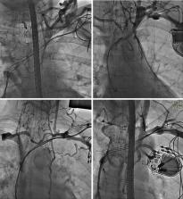
          Lead-dependent venous occlusion may impede the insertion of a central venous access device (CVAD). The aim of this retrospective, cohort study was to assess the chance of implantation of CVAD in patients with cardiac implantable electronic devices (CIEDs).

          Methods

          We reviewed and analyzed 3,075 venograms of patients with CIEDs undergoing transvenous lead extraction (TLE) between June 2008 and July 2021. Relationship between venous patency and the chance of CVAD placement was estimated.

          Results

          In 2,318 (75.38%) patients, venography showed no potential obstacles to venous port implantation on the ipsilateral side. In patients with leads on the left side, significant narrowing more often affected the subclavian vein than the brachiocephalic vein [1,595 (55.29%) vs. 830 (28.63%), respectively] or the superior vena cava (SVC) [21 (0.73%) cases]. Furthermore, the subclavian and brachiocephalic veins on the opposite side were also narrowed [35 (2.35%) and 27 (1.24%), respectively]. The chances of port insertion were assessed as easy on CIED side or opposite side in 2,318 (75.38%) and 2,291 (97.91%) patients, respectively), as difficult insertion/questionable performance in 246 (8.00%) and 22 (0.94% patients) and doubtful or impossible insertion/questionable performance in 511 (16.62%)/27 (1.15%) patients with CIED.

          Conclusions

          (I) Varying degrees of lead-dependent venous obstruction (LDVO) is a frequent finding in patients with CIEDs; (II) the major thoracic veins on the opposite side of the chest may also be significantly narrowed; (III) venography should be considered before attempted CVAD insertion in patients with long lead dwell times or in patients after CIED removal, including planned contralateral port placement.

          Related collections

          Most cited references44

          • Record: found
          • Abstract: found
          • Article: found
          Is Open Access

          Ultrasound-guided central venous catheter placement: a structured review and recommendations for clinical practice

          The use of ultrasound (US) has been proposed to reduce the number of complications and to increase the safety and quality of central venous catheter (CVC) placement. In this review, we describe the rationale for the use of US during CVC placement, the basic principles of this technique, and the current evidence and existing guidelines for its use. In addition, we recommend a structured approach for US-guided central venous access for clinical practice. Static and real-time US can be used to visualize the anatomy and patency of the target vein in a short-axis and a long-axis view. US-guided needle advancement can be performed in an "out-of-plane" and an "in-plane" technique. There is clear evidence that US offers gains in safety and quality during CVC placement in the internal jugular vein. For the subclavian and femoral veins, US offers small gains in safety and quality. Based on the available evidence from clinical studies, several guidelines from medical societies strongly recommend the use of US for CVC placement in the internal jugular vein. Data from survey studies show that there is still a gap between the existing evidence and guidelines and the use of US in clinical practice. For clinical practice, we recommend a six-step systematic approach for US-guided central venous access that includes assessing the target vein (anatomy and vessel localization, vessel patency), using real-time US guidance for puncture of the vein, and confirming the correct needle, wire, and catheter position in the vein. To achieve the best skill level for CVC placement the knowledge from anatomic landmark techniques and the knowledge from US-guided CVC placement need to be combined and integrated.
            Bookmark
            • Record: found
            • Abstract: found
            • Article: not found

            Incidence and risk factors of early venous thrombosis associated with permanent pacemaker leads.

            Pacemaker lead implantation can cause thrombosis, which can be associated with serious local morbidity and complicated by pulmonary embolism. Few reliable estimates of the incidence of thrombosis have been reported. The contribution of established risk factors to venous thrombosis in patients with implanted pacemaker leads is unknown. One hundred forty-five consecutive patients n = 145) underwent routine clinical and Doppler ultrasound evaluation for thrombosis before and 3, 6, and 12 months after lead implantation. Established risk factors for venous thrombosis were assessed in detail for all patients. Clinical outcome, including clinically manifest thrombosis, pulmonary embolism, associated pacemaker lead infection, complicated reinterventions, and death, was evaluated. Thrombosis was observed in 34 (23%) of 145 patients. Thrombosis did not cause any signs or symptoms in 31 patients but resulted in overt clinical symptoms in 3 patients. The absence of anticoagulant therapy, use of hormone therapy, and a personal history of venous thrombosis were associated with an increased risk of thrombosis. The risk of thrombosis increased in the presence of multiple pacemaker leads compared to a single lead. Established risk factors for venous thrombosis and the presence of multiple pacemaker leads contribute substantially to the occurrence of thrombosis associated with permanent pacemaker leads. Risk factor assessment prior to implantation may be useful for identifying patients at risk for thrombotic complications. Preventive management in these patients is warranted.
              Bookmark
              • Record: found
              • Abstract: found
              • Article: not found

              Incidence of venous obstruction following insertion of an implantable cardioverter defibrillator. A study of systematic contrast venography on patients presenting for their first elective ICD generator replacement.

              The number of implantable cardioverter defibrillator (ICD) implantations, as well as follow-up procedures such as generator exchanges, lead revisions and lead system upgrades, is ever-increasing. Lead revisions and implantation of additional leads require venous access at the site of the previous ICD implantation. The aim of our study was therefore to evaluate the incidence of venous obstruction after chronic transvenous ICD system implantation. One hundred and five consecutive patients admitted for their first elective ICD generator replacement were included. All patients underwent bilateral contrast venography and the images were analyzed by two attending radiologists. Venous obstruction was classified as moderate stenosis (50-75% diameter reduction), severe stenosis (>75%) or total occlusion. Venous obstruction of various degrees was found in 25% of the patients. Complete occlusion was found in 9%, severe stenosis in 6% and moderate stenosis in 10% of the patients. The incidence of venous obstruction was increased in patients with a pacemaker prior to the initial ICD system implantation (67%). No difference was found in patients with a single defibrillator lead compared with patients who had an additional superior vena cava (SVC) shocking coil. However, the presence of a second shocking coil in the SVC incorporated in a single ICD lead was associated with an increased incidence of venous obstruction. No difference was found between silicone and polyurethane insulated leads. This study shows that venous obstruction occurs relatively frequently after ICD implantation. Therefore, contrast venography should always be obtained if malfunction of a preexistent lead is suspected or a system upgrade is considered.
                Bookmark

                Author and article information

                Journal
                Cardiovasc Diagn Ther
                Cardiovasc Diagn Ther
                CDT
                Cardiovascular Diagnosis and Therapy
                AME Publishing Company
                2223-3652
                2223-3660
                27 October 2023
                15 December 2023
                : 13
                : 6
                : 1068-1079
                Affiliations
                [1 ]deptDepartment of Cardiac Surgery , Medical University of Lublin , Lublin, Poland;
                [2 ]deptDepartment of Physiology, Pathophysiology and Clinical Immunology, Institute of Medical Sciences , Jan Kochanowski University , Kielce, Poland;
                [3 ]deptDepartment of Cardiac Surgery , Świętokrzyskie Cardiology Center , Kielce, Poland;
                [4 ]2nd Department of Cardiology, Faculty of Medical Science in Zabrze, Medical University of Silesia in Katowice, Zabrze, Poland;
                [5 ]Department of Cardiology, The Pope John Paul II Province Hospital of Zamość, Zamość , Poland;
                [6 ]Department of Cardiac Surgery, The Pope John Paul II Province Hospital of Zamość, Zamość , Poland;
                [7 ]deptDepartment of Cardiology , Medical University of Lublin , Lublin, Poland
                Author notes

                Contributions: (I) Conception and design: M Czajkowski, A Kutarski; (II) Administrative support: A Polewczyk; (III) Provision of study materials or patients: Ł Tułecki, P Stefańczyk, A Kutarski; (IV) Collection and assembly of data: D Nowosielecka, Ł Tułecki, P Stefańczyk, A Kutarski; (V) Data analysis and interpretation: W Jacheć, M Czajkowski, A Polewczyk, A Kutarski; (VI) Manuscript writing: All authors; (VII) Final approval of manuscript: All authors.

                Correspondence to: Anna Polewczyk, MD, PhD. Department of Physiology, Pathophysiology and Clinical Immunology, Institute of Medical Sciences, Jan Kochanowski University, 5, Żeromskiego St. 25-369 Kielce, Poland; Department of Cardiac Surgery, Świętokrzyskie Cardiology Center, Kielce, Poland. Email: annapolewczyk@ 123456wp.pl .
                [^]

                ORCID: 0000-0002-6632-6551.

                Article
                cdt-13-06-1068
                10.21037/cdt-23-104
                10753236
                b0d7b65d-e64f-4cd8-8397-c751368f653b
                2023 Cardiovascular Diagnosis and Therapy. All rights reserved.

                Open Access Statement: This is an Open Access article distributed in accordance with the Creative Commons Attribution-NonCommercial-NoDerivs 4.0 International License (CC BY-NC-ND 4.0), which permits the non-commercial replication and distribution of the article with the strict proviso that no changes or edits are made and the original work is properly cited (including links to both the formal publication through the relevant DOI and the license). See: https://creativecommons.org/licenses/by-nc-nd/4.0.

                History
                : 13 March 2023
                : 07 August 2023
                Categories
                Original Article

                endocardial leads,venography,lead-related venous obstruction,venous port implantation,venous port complications

                Comments

                Comment on this article