8
views
0
recommends
+1 Recommend
0 collections
    0
    shares
      • Record: found
      • Abstract: found
      • Article: found
      Is Open Access

      Commentary: Clinical and biometric characteristics of pediatric eyes with nanophthalmos

      article-commentary

      Read this article at

      Bookmark
          There is no author summary for this article yet. Authors can add summaries to their articles on ScienceOpen to make them more accessible to a non-specialist audience.

          Abstract

          The development of the eye starts as early as three weeks of gestational age.[1] It involves the differentiation of specific cells from neuroepithelium and mesenchyme into various ocular tissues, which perform particular functions aiding in vision. Mutations in specific genes encoding for the development of the eye, such as PAX6, MRFP, TMEM98, BEST1, lead to developmental abnormalities in the eye.[2] These abnormalities can be as minimal as an isolated iris coloboma to gross defects like microphthalmos. Microphthalmos is a decrease in the eye’s size compared to age-matched population. Nanophthalmos is a clinical variant of microphthalmos in which the anterior and posterior segments of the eye stop developing without any other functional or structural abnormality.[3] Nanophthalmos is considered a rare/orphan disease by the Orphan Drug Act of 1983, which means the prevalence is considerably low in the general population to provide a minuscule financial incentive for the private sector to make and market new medications to treat or prevent it. The diagnosis and classification of this relatively rare disease is thus understudied. However, it is essential to study nanophthalmos as it provides us with information about the embryological development of the eye. The article in the present issue of the journal details the characteristics of 40 patients with nanophthalmos, comparing them with the normal population.[4] It is crucial to understand that definition of nanophthalmos cannot be simplified to <20.5 mm, as used in the present article, for all age groups. Similarly, a blanket value of less than <16 years with no age range and an axial length cutoff value of 17 mm doesn’t provide useful clinical information. There are varying classifications of nanophthalmos, but pediatric definitions are not clear.[5 6] Axial length less than two standard deviations for that age with other coexisting factors of high hyperopia (>+7 D), a small anterior chamber with a normal/thicker lens and steep, smaller corneas (<11 mm), as well as retinochoroidal thickening are 
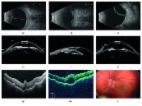
essential in classifying a patient as nanophthalmic. Hence, it is necessary to measure the biometric parameters, including the retino-choroido-scleral thickness and the LT/ACD or LT/AL ratio when diagnosing nanophthalmos.[7] Identifying these factors is essential as almost half of those with higher ratios are at risk of developing glaucoma.[7] Clinical features of nanophthalmos play an essential role in diagnosing the disease and differentiating it from posterior microphthalmos with normal anterior segment dimensions (hence not at risk for glaucoma) but more retinal complications.[3 8] A combination of increased scleral thickness and abnormal collagen is hypothesized to impair vortex venous drainage and reduce the transscleral flow of proteins in these patients. Recent genetic studies underlie a strong familial basis of nanophthalmos, with five genes (MFRP, TMEM98, PRSS56, BEST1, and CRB1) being implicated.[3] Nanophthalmos is an easily missed diagnosis due to the complexity of its evaluation and classification, making its diagnosis challenging. Historically speaking, the anatomy and histology of ocular structures in such cases were too complicated to evaluate with the available resources. With the advent of optical coherence tomography (OCT), it is easier to document and record findings in these patients. There is minimal source available on nanophthalmic pediatric patients. Pediatric ophthalmologists should also be well versed with this condition, which will help in the early diagnosis and treatment of associated complications like hyperopia (and the resulting amblyopia), angle-closure glaucoma, and uveal effusion syndrome.

          Related collections

          Most cited references8

          • Record: found
          • Abstract: found
          • Article: found
          Is Open Access

          Nanophthalmos: A Review of the Clinical Spectrum and Genetics

          Nanophthalmos is a clinical spectrum of disorders with a phenotypically small but structurally normal eye. These disorders present significant clinical challenges to ophthalmologists due to a high rate of secondary angle-closure glaucoma, spontaneous choroidal effusions, and perioperative complications with cataract and retinal surgeries. Nanophthalmos may present as a sporadic or familial disorder, with autosomal-dominant or recessive inheritance. To date, five genes (i.e., MFRP, TMEM98, PRSS56, BEST1, and CRB1) and two loci have been implicated in familial forms of nanophthalmos. Here, we review the definition of nanophthalmos, the clinical and pathogenic features of the condition, and the genetics of this disorder.
            Bookmark
            • Record: found
            • Abstract: found
            • Article: not found

            Management of glaucoma in patients with nanophthalmos.

            In this retrospective study, we aimed to evaluate the results of glaucoma surgery in patients with nanophthalmos. Twenty-eight bilateral nanophthalmic patients, of whom 20 patients underwent trabeculectomy+Mitomycin-C (MMC)+inferior sclerotomy between 1996 and 2004, were included in this study. Records of patients with nanophthalmos were reviewed. Intraocular pressure (IOP), glaucoma medications, surgical success, visual acuity and complications were analyzed. The mean IOP was 34.6+/-5.3 mm Hg preoperatively. At the final follow-up visit, the mean IOP dropped to 21.41+/-7.34 mm Hg (P<0.05). The cumulative probability of success was 85% at 1 year, 78.5% at 2 years, 76.9% at 3 years, 70.6% at 4 years and 47% at 5 years after surgery. Visual acuity decreased in 13 (65%) patients but no eye lost vision. Sequels of choroidal detachment (five patients 25%) and retinal folds (four patients 20%) were the most frequent reason for visual decrease. Uveal effusion (10 patients 50%) and cataract formation (seven patients 35%) were major late postoperative complications. Results indicate that trabeculectomy+MMC+inferior sclerotomy procedure was effective and safe for glaucoma control in patients with nanophthalmos, but uveal effusion is a major problem and cataract surgery is expected in the long run.
              Bookmark
              • Record: found
              • Abstract: found
              • Article: not found

              Cataract surgery in patients with nanophthalmos: results and complications.

              To evaluate the results and complications of cataract surgery in patients with nanophthalmos. University hospital practice. The records of consecutive patients with nanophthalmos who had cataract surgery from 1978 through 2002 were reviewed for ocular diagnoses, corneal diameter, keratometry, axial length, retinal-choroidal-scleral thickness determined by echography, ocular surgeries, visual acuity, and complications. Eight patients (6 women, 2 men) with a mean age of 59 years were reviewed. Four patients were not previously diagnosed with nanophthalmos; increased retinal-choroidal-scleral thickness (mean 2.41 mm) confirmed the diagnosis. Twelve eyes had cataract extraction with posterior chamber intraocular lens (IOL) implantation, 11 by phacoemulsification and 1 by extracapsular cataract extraction, and 4 eyes had lamellar scleral resections. Additional surgeries included glaucoma laser treatment (8 eyes), cyclocryotherapy (2 eyes), trabeculectomy with scleral resection (1 eye), trabeculectomy combined with phacoemulsification (1 eye), and neodymium:YAG laser capsulotomy (4 eyes). No eye lost vision; however, complications included severe iritis, broken IOL haptic with vitreous loss, posterior capsule opacity, choroidal hemorrhage, phthisis, and aqueous misdirection. Results indicate that echography should be used to assess retinal-choroidal-scleral thickness in eyes that are hyperopic and at risk for narrow-angle glaucoma. Thickening may confirm the diagnosis of nanophthalmos and allow careful preoperative assessment and appropriate operative procedures in these high-risk eyes. With advances in cataract, glaucoma, and uveal effusion treatments, surgical results in patients with nanophthalmos are improving.
                Bookmark

                Author and article information

                Journal
                Indian J Ophthalmol
                Indian J Ophthalmol
                IJO
                Indian J Ophthalmol
                Indian Journal of Ophthalmology
                Wolters Kluwer - Medknow (India )
                0301-4738
                1998-3689
                July 2022
                July 2022
                : 70
                : 7
                : 2447-2448
                Affiliations
                [1]Advanced Eye Centre, Post Graduate Institute of Medical Education and Research, Chandigarh, India
                Author notes
                Correspondence to: Dr. Savleen Kaur, Assistant Professor, Advanced Eye Centre (Department of Ophthalmology), Post Graduate Institute of Medical Education and Research, Chandigarh, India. E-mail: mailsavleen@ 123456gmail.com
                Article
                IJO-70-2447
                10.4103/ijo.IJO_844_22
                9426170
                35791129
                af7f612d-754c-457e-b0d0-d1577b6ea553
                Copyright: © 2022 Indian Journal of Ophthalmology

                This is an open access journal, and articles are distributed under the terms of the Creative Commons Attribution-NonCommercial-ShareAlike 4.0 License, which allows others to remix, tweak, and build upon the work non-commercially, as long as appropriate credit is given and the new creations are licensed under the identical terms.

                History
                Categories
                Commentary

                Ophthalmology & Optometry
                Ophthalmology & Optometry

                Comments

                Comment on this article