36
views
0
recommends
+1 Recommend
0 collections
    0
    shares
      • Record: found
      • Abstract: found
      • Article: found
      Is Open Access

      Thyroid hormone signaling specifies cone subtypes in human retinal organoids

      research-article

      Read this article at

      Bookmark
          There is no author summary for this article yet. Authors can add summaries to their articles on ScienceOpen to make them more accessible to a non-specialist audience.

          Abstract

          INTRODUCTION:

          Cone photoreceptors in the human retina enable daytime, color, and high-acuity vision. The three subtypes of human cones are defined by the visual pigment that they express: blue-opsin (short wavelength; S), green-opsin (medium wavelength; M), or red-opsin (long wavelength; L). Mutations that affect opsin expression or function cause various forms of color blindness and retinal degeneration.

          RATIONALE:

          Our current understanding of the vertebrate eye has been derived primarily from the study of model organisms. We studied the human retina to understand the developmental mechanisms that generate the mosaic of mutually exclusive cone subtypes. Specification of human cones occurs in a two-step process. First, a decision occurs between S versus L/M cone fates. If the L/M fate is chosen, a subsequent choice is made between expression of L- or M-opsin. To determine the mechanism that controls the first decision between S and L/M cone fates, we studied human retinal organoids derived from stem cells.

          RESULTS:

          We found that human organoids and retinas have similar distributions, gene expression profiles, and morphologies of cone subtypes. During development, S cones are specified first, followed by L/M cones. This temporal switch from specification of S cones to generation of L/M cones is controlled by thyroid hormone (TH) signaling. In retinal organoids that lacked thyroid hormone receptor β ( Thrβ), all cones developed into the S subtype. Thrβ binds with high affinity to triiodothyronine (T3), the more active form of TH, to regulate gene expression. We observed that addition of T3 early during development induced L/M fate in nearly all cones. Thus, TH signaling through Thrβ is necessary and sufficient to induce L/M cone fate and suppress S fate. TH exists largely in two states: thyroxine (T4), the most abundant circulating form of TH, and T3, which binds TH receptors with high affinity. We hypothesized that the retina itself could modulate TH levels to control subtype fates. We found that deiodinase 3 ( DIO3), an enzyme that degrades both T3 and T4, was expressed early in organoid and retina development. Conversely, deiodinase 2 ( DIO2), an enzyme that converts T4 to active T3, as well as TH carriers and transporters, were expressed later in development. Temporally dynamic expression of TH-degrading and -activating proteins supports a model in which the retina itself controls TH levels, ensuring low TH signaling early to specify S cones and high TH signaling later in development to produce L/M cones.

          CONCLUSION:

          Studies of model organisms and human epidemiology often generate hypotheses about human biology that cannot be studied in humans. Organoids provide a system to determine the mechanisms of human development, enabling direct testing of hypotheses in developing human tissue. Our studies identify temporal regulation of TH signaling as a mechanism that controls cone subtype specification in humans. Consistent with our findings, preterm human infants with low T3 and T4 have an increased incidence of color vision defects. Moreover, our identification of a mechanism that generates one cone subtype while suppressing the other, coupled with successful transplantation and incorporation of stem cell-derived photoreceptors in mice, suggests that the promise of therapies to treat human diseases such as color blindness, retinitis pigmentosa, and macular degeneration will be achieved in the near future. ■

          Graphical Abstract

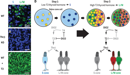
          Temporally regulated TH signaling specifies cone subtypes. ( A) Embryonic stem cell-derived human retinal organoids [wild type (WT)] generate S and L/M cones. Blue, S-opsin; green, L/M-opsin. ( B) Organoids that lack thyroid hormone receptor β ( Thrβ KO) generate all S cones. ( C) Early activation of TH signaling (WT + T3) specifies nearly all L/M cones. ( D) TH-degrading enzymes (such as DIO3) expressed early in development lower TH and promote S fate, whereas TH-activating regulators (such as DIO2) expressed later promote L/M fate.

          Summary

          The mechanisms underlying specification of neuronal subtypes within the human nervous system are largely unknown. The blue (S), green (M), and red (L) cones of the retina enable high-acuity daytime and color vision. To determine the mechanism that controls S versus L/M fates, we studied the differentiation of human retinal organoids. Organoids and retinas have similar distributions, expression profiles, and morphologies of cone subtypes. S cones are specified first, followed by L/M cones, and thyroid hormone signaling controls this temporal switch. Dynamic expression of thyroid hormone–degrading and –activating proteins within the retina ensures low signaling early to specify S cones and high signaling late to produce L/M cones. This work establishes organoids as a model for determining mechanisms of human development with promising utility for therapeutics and vision repair.

          Related collections

          Most cited references48

          • Record: found
          • Abstract: found
          • Article: not found

          Biochemistry, cellular and molecular biology, and physiological roles of the iodothyronine selenodeiodinases.

          The goal of this review is to place the exciting advances that have occurred in our understanding of the molecular biology of the types 1, 2, and 3 (D1, D2, and D3, respectively) iodothyronine deiodinases into a biochemical and physiological context. We review new data regarding the mechanism of selenoprotein synthesis, the molecular and cellular biological properties of the individual deiodinases, including gene structure, mRNA and protein characteristics, tissue distribution, subcellular localization and topology, enzymatic properties, structure-activity relationships, and regulation of synthesis, inactivation, and degradation. These provide the background for a discussion of their role in thyroid physiology in humans and other vertebrates, including evidence that D2 plays a significant role in human plasma T(3) production. We discuss the pathological role of D3 overexpression causing "consumptive hypothyroidism" as well as our current understanding of the pathophysiology of iodothyronine deiodination during illness and amiodarone therapy. Finally, we review the new insights from analysis of mice with targeted disruption of the Dio2 gene and overexpression of D2 in the myocardium.
            Bookmark
            • Record: found
            • Abstract: found
            • Article: not found

            Transplantation of human embryonic stem cell-derived photoreceptors restores some visual function in Crx-deficient mice.

            Some of the most common causes of blindness involve the degeneration of photoreceptors in the neural retina; photoreceptor replacement therapy might restore some vision in these individuals. Embryonic stem cells (ESCs) could, in principle, provide a source of photoreceptors to repair the retina. We have previously shown that retinal progenitors can be efficiently derived from human ESCs. We now show that retinal cells derived from human ESCs will migrate into mouse retinas following intraocular injection, settle into the appropriate layers, and express markers for differentiated cells, including both rod and cone photoreceptor cells. After transplantation of the cells into the subretinal space of adult Crx(-/-) mice (a model of Leber's Congenital Amaurosis), the hESC-derived retinal cells differentiate into functional photoreceptors and restore light responses to the animals. These results demonstrate that hESCs can, in principle, be used for photoreceptor replacement therapies.
              Bookmark
              • Record: found
              • Abstract: found
              • Article: not found

              Crx, a novel Otx-like paired-homeodomain protein, binds to and transactivates photoreceptor cell-specific genes.

              The otd/Otx gene family encodes paired-like homeodomain proteins that are involved in the regulation of anterior head structure and sensory organ development. Using the yeast one-hybrid screen with a bait containing the Ret 4 site from the bovine rhodopsin promoter, we have cloned a new member of the family, Crx (Cone rod homeobox). Crx encodes a 299 amino acid residue protein with a paired-like homeodomain near its N terminus. In the adult, it is expressed predominantly in photoreceptors and pinealocytes. In the developing mouse retina, it is expressed by embryonic day 12.5 (E12.5). Recombinant Crx binds in vitro not only to the Ret 4 site but also to the Ret 1 and BAT-1 sites. In transient transfection studies, Crx transactivates rhodopsin promoter-reporter constructs. Its activity is synergistic with that of Nrl. Crx also binds to and transactivates the genes for several other photoreceptor cell-specific proteins (interphotoreceptor retinoid-binding protein, beta-phosphodiesterase, and arrestin). Human Crx maps to chromosome 19q13.3, the site of a cone rod dystrophy (CORDII). These studies implicate Crx as a potentially important regulator of photoreceptor cell development and gene expression and also identify it as a candidate gene for CORDII and other retinal diseases.
                Bookmark

                Author and article information

                Journal
                0404511
                7473
                Science
                Science
                Science (New York, N.Y.)
                0036-8075
                1095-9203
                20 October 2018
                12 October 2018
                12 May 2019
                : 362
                : 6411
                : eaau6348
                Affiliations
                [1 ]Department of Biology, Johns Hopkins University, 3400 N. Charles Street, Baltimore, MD 21218, USA.
                [2 ]Wilmer Eye Institute, Johns Hopkins University School of Medicine, Baltimore, MD 21287, USA.
                [3 ]Shiley Eye Institute, University of California, San Diego, La Jolla, CA 92093, USA.
                [4 ]National Institute of Mental Health, National Institutes of Health, Bethesda, MD 20892, USA.
                [5 ]Department of Computer Science, Johns Hopkins University, 3400 N. Charles Street, Baltimore, MD 21218, USA.
                [6 ]Department of Molecular Biology and Genetics, Johns Hopkins University School of Medicine, Baltimore, MD 21287, USA.
                [7 ]Department of Neuroscience, Johns Hopkins University School of Medicine, Baltimore, MD 21287, USA.
                [8 ]Institute of Genetic Medicine, Johns Hopkins University School of Medicine, Baltimore, MD 21287, USA.
                Author notes
                [* ]Corresponding author. robertjohnston@ 123456jhu.edu

                Author Contributions: K.C.E.: Conception, data acquisition, new reagent contribution, data analysis, and data interpretation; drafted and revised manuscript. S.E.H.: Data acquisition and data interpretation. K.A.H.: Data acquisition, data analysis, and data interpretation. B.B.: Data analysis and data interpretation. P.-W.Z.: New reagent contribution. X.C.: New reagent contribution. V.M.S.: New reagent contribution. D.S.W.: New reagent contribution. S.H.: Data interpretation. J.T.: Data analysis and data interpretation. K.W.: Data acquisition and new reagent contribution. D.J.Z.: Data acquisition and new reagent contribution. R.J.J.: Conception and data interpretation; drafted and revised manuscript.

                Article
                NIHMS993285
                10.1126/science.aau6348
                6249681
                30309916
                af6279a0-a616-4866-9eae-a920227319be

                exclusive licensee American Association for the Advancement of Science. http://creativecommons.org/licenses/by/4.0/

                History
                Categories
                Article

                Uncategorized
                Uncategorized

                Comments

                Comment on this article