34
views
0
recommends
+1 Recommend
0 collections
    0
    shares
      • Record: found
      • Abstract: found
      • Article: not found

      A Histopathology Study of Caspian Seal ( Pusa caspica) (Phocidae, Mammalia) Liver Infected with Trematode, Pseudamphistomum truncatum (Rudolphi, 1819) (Opisthorchidae, Trematoda)

      research-article

      Read this article at

      ScienceOpenPMC
      Bookmark
          There is no author summary for this article yet. Authors can add summaries to their articles on ScienceOpen to make them more accessible to a non-specialist audience.

          Abstract

          Background

          Main objective of this study was to investigate the invasive activity of the liver fluke, Pseudamphistomom truncatum against the Caspian seal ( Pusa caspica) and was exemplified at the gross, light microscopy (LM) and electron microscopy (EM) levels.

          Methods

          The study was done on a freshly dead Caspian Seal in the southern coast of Caspian Sea. The checked Caspian seal probably being died of canine distemper virus and was found host to numerous parasites of four helminth species.

          Results

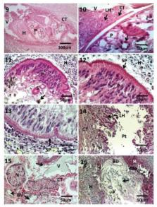
          P. truncatum caused edematous foci on the surface of the liver with prominent fluid accumulation. Sections of the liver viewed with LM had multiple necrotic areas with extensive hemorrhaging and disorganized hepatic lobules. Granulocytes and invasion of connective tissue were prominent. Whole worms were visible with invasive pathways through the host tissue. Damage to both hepatic ducts and blood vessels were prominent. At the EM level, organelles within the impacted hepatocytes were disorganized as exemplified by the cristae of the mitochondria and the endoplasmic reticulum. Parasite eggs were scattered throughout the tissue.

          Conclusion

          It was shown that this trematode can be very pathogenic to Caspian Seal and as this only mammal of Caspian Sea is an endangered species; this needs more investigation toward control or possible treatment of this helminth.

          Related collections

          Most cited references25

          • Record: found
          • Abstract: found
          • Article: not found

          Mass die-Off of Caspian seals caused by canine distemper virus.

          Thousands of Caspian seals (Phoca caspica) died in the Caspian Sea from April to August 2000. Lesions characteristic of morbillivirus infection were found in tissue specimens from dead seals. Canine distemper virus infection was identified by serologic examination, reverse transcriptase- polymerase chain reaction, and sequencing of selected P gene fragments. These results implicate canine distemper virus infection as the primary cause of death.
            Bookmark
            • Record: found
            • Abstract: not found
            • Book: not found

            Theory and Practice of Histological Techniques

              Bookmark
              • Record: found
              • Abstract: found
              • Article: not found

              The morphology of an unique population of Corynosoma strumosum (Acanthocephala, Polymorphidae) from the Caspian seal, Pusa caspica, in the land-locked Caspian Sea using SEM, with special notes on histopathology

              Over 700 specimens of Corynosoma strumosum (Rudolphi, 1802) Lühe, 1904 were collected from one young male Caspian seal, Pusa caspica (Gmelin) in the southern land-locked Caspian Sea in April, 2009. Collected worms showed consistent variations from those reported by other observers using light microscopy especially in proboscis hook and trunk spine patterns. SEM images revealed many features that have not been previously reported including the shape and distribution of trunk spines, dorsoventral differences in proboscis hooks and their organization, the baldness of anterior proboscis, the rough egg topography, epidermal micropores, and variations in the female gonopore. This isolated population of C. strumosum from the land-locked Caspian Sea is distinguished from others reported from open waters elsewhere by the distribution of trunk spines, consistently smaller size of trunk and testes, larger eggs, and fewer proboscis hooks. Histopathological sections reveal the invasive path of worms in host tissue with damage to intestinal villi and worm encapsulation. Information obtained from SEM studies and histopathological sections is reported for the first time.
                Bookmark

                Author and article information

                Journal
                Iran J Parasitol
                Iran J Parasitol
                IJPA
                Iranian Journal of Parasitology
                Tehran University of Medical Sciences
                1735-7020
                2008-238X
                Apr-Jun 2014
                : 9
                : 2
                : 266-275
                Affiliations
                [1 ]Dept. of Biology, Brigham Young University, Provo, Utah, USA
                [2 ]Dept. of Biodiversity (Zoology), University of Limpopo, Turfloop Campus, Sovenga 0727, Polokwane, South Africa
                [3 ]College of Women for Arts, Science and Education, Ain Shams University, Cairo, Egypt and Brigham Young University, Provo, Utah, USA
                Author notes
                Article
                4386049
                adc47faa-53b4-4efd-abd8-16aa462cfa52
                Copyright: © Iranian Journal of Parasitology & Tehran University of Medical Sciences

                This work is licensed under a Creative Commons Attribution-NonCommercial 3.0 Unported License which allows users to read, copy, distribute and make derivative works for non-commercial purposes from the material, as long as the author of the original work is cited properly.

                History
                : 24 September 2013
                : 11 January 2014
                Categories
                Short Communication

                Parasitology
                caspian seal,iran,p. truncatum,pathology,liver
                Parasitology
                caspian seal, iran, p. truncatum, pathology, liver

                Comments

                Comment on this article