14
views
0
recommends
+1 Recommend
0 collections
    0
    shares
      • Record: found
      • Abstract: found
      • Article: found
      Is Open Access

      Kinetic modeling and parametric imaging with dynamic PET for oncological applications: general considerations, current clinical applications, and future perspectives

      review-article

      Read this article at

      Bookmark
          There is no author summary for this article yet. Authors can add summaries to their articles on ScienceOpen to make them more accessible to a non-specialist audience.

          Abstract

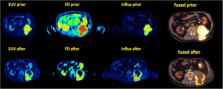
          Dynamic PET (dPET) studies have been used until now primarily within research purposes. Although it is generally accepted that the information provided by dPET is superior to that of conventional static PET acquisitions acquired usually 60 min post injection of the radiotracer, the duration of dynamic protocols, the limited axial field of view (FOV) of current generation clinical PET systems covering a relatively small axial extent of the human body for a dynamic measurement, and the complexity of data evaluation have hampered its implementation into clinical routine. However, the development of new-generation PET/CT scanners with an extended FOV as well as of more sophisticated evaluation software packages that offer better segmentation algorithms, automatic retrieval of the arterial input function, and automatic calculation of parametric imaging, in combination with dedicated shorter dynamic protocols, will facilitate the wider use of dPET. This is expected to aid in oncological diagnostics and therapy assessment. The aim of this review is to present some general considerations about dPET analysis in oncology by means of kinetic modeling, based on compartmental and noncompartmental approaches, and parametric imaging. Moreover, the current clinical applications and future perspectives of the modality are outlined.

          Related collections

          Most cited references99

          • Record: found
          • Abstract: found
          • Article: not found

          From RECIST to PERCIST: Evolving Considerations for PET response criteria in solid tumors.

          The purpose of this article is to review the status and limitations of anatomic tumor response metrics including the World Health Organization (WHO) criteria, the Response Evaluation Criteria in Solid Tumors (RECIST), and RECIST 1.1. This article also reviews qualitative and quantitative approaches to metabolic tumor response assessment with (18)F-FDG PET and proposes a draft framework for PET Response Criteria in Solid Tumors (PERCIST), version 1.0. PubMed searches, including searches for the terms RECIST, positron, WHO, FDG, cancer (including specific types), treatment response, region of interest, and derivative references, were performed. Abstracts and articles judged most relevant to the goals of this report were reviewed with emphasis on limitations and strengths of the anatomic and PET approaches to treatment response assessment. On the basis of these data and the authors' experience, draft criteria were formulated for PET tumor response to treatment. Approximately 3,000 potentially relevant references were screened. Anatomic imaging alone using standard WHO, RECIST, and RECIST 1.1 criteria is widely applied but still has limitations in response assessments. For example, despite effective treatment, changes in tumor size can be minimal in tumors such as lymphomas, sarcoma, hepatomas, mesothelioma, and gastrointestinal stromal tumor. CT tumor density, contrast enhancement, or MRI characteristics appear more informative than size but are not yet routinely applied. RECIST criteria may show progression of tumor more slowly than WHO criteria. RECIST 1.1 criteria (assessing a maximum of 5 tumor foci, vs. 10 in RECIST) result in a higher complete response rate than the original RECIST criteria, at least in lymph nodes. Variability appears greater in assessing progression than in assessing response. Qualitative and quantitative approaches to (18)F-FDG PET response assessment have been applied and require a consistent PET methodology to allow quantitative assessments. Statistically significant changes in tumor standardized uptake value (SUV) occur in careful test-retest studies of high-SUV tumors, with a change of 20% in SUV of a region 1 cm or larger in diameter; however, medically relevant beneficial changes are often associated with a 30% or greater decline. The more extensive the therapy, the greater the decline in SUV with most effective treatments. Important components of the proposed PERCIST criteria include assessing normal reference tissue values in a 3-cm-diameter region of interest in the liver, using a consistent PET protocol, using a fixed small region of interest about 1 cm(3) in volume (1.2-cm diameter) in the most active region of metabolically active tumors to minimize statistical variability, assessing tumor size, treating SUV lean measurements in the 1 (up to 5 optional) most metabolically active tumor focus as a continuous variable, requiring a 30% decline in SUV for "response," and deferring to RECIST 1.1 in cases that do not have (18)F-FDG avidity or are technically unsuitable. Criteria to define progression of tumor-absent new lesions are uncertain but are proposed. Anatomic imaging alone using standard WHO, RECIST, and RECIST 1.1 criteria have limitations, particularly in assessing the activity of newer cancer therapies that stabilize disease, whereas (18)F-FDG PET appears particularly valuable in such cases. The proposed PERCIST 1.0 criteria should serve as a starting point for use in clinical trials and in structured quantitative clinical reporting. Undoubtedly, subsequent revisions and enhancements will be required as validation studies are undertaken in varying diseases and treatments.
            Bookmark
            • Record: found
            • Abstract: found
            • Article: not found

            Measurement of clinical and subclinical tumour response using [18F]-fluorodeoxyglucose and positron emission tomography: review and 1999 EORTC recommendations. European Organization for Research and Treatment of Cancer (EORTC) PET Study Group.

            [18F]-fluorodeoxyglucose ([18F]-FDG) uptake is enhanced in most malignant tumours which in turn can be measured using positron emission tomography (PET). A number of small clinical trials have indicated that quantification of the change in tumour [18F]-FDG uptake may provide an early, sensitive, pharmacodynamic marker of the tumoricidal effect of anticancer drugs. This may allow for the introduction of subclinical response for anticancer drug evaluation in early clinical trials and improvements in patient management. For comparison of results from smaller clinical trials and larger-scale multicentre trials a consensus is desirable for: (i) common measurement criteria; and (ii) reporting of alterations in [18F]-FDG uptake with treatment. This paper summarises the current status of the technique and recommendations on the measurement of [18F]-FDG uptake for tumour response monitoring from a consensus meeting of the European Organization for Research and Treatment of Cancer (EORTC) PET study group held in Brussels in February 1998 and confirmed at a subsequent meeting in March 1999.
              Bookmark
              • Record: found
              • Abstract: found
              • Article: not found

              First Human Imaging Studies with the EXPLORER Total-Body PET Scanner*

              Within the EXPLORER Consortium, the construction of the world's first total-body PET/CT scanner has recently been completed. The 194-cm axial field of view of the EXPLORER PET/CT scanner is sufficient to cover, for the first time, the entire human adult body in a single acquisition in more than 99% of the population and allows total-body pharmacokinetic studies with frame durations as short as 1 s. The large increase in sensitivity arising from total-body coverage as well as increased solid angle for detection at any point within the body allows whole-body 18F-FDG PET studies to be acquired with unprecedented count density, improving the signal-to-noise ratio of the resulting images. Alternatively, the sensitivity gain can be used to acquire diagnostic PET images with very small amounts of activity in the field of view (25 MBq, 0.7 mCi or less), with very short acquisition times (∼1 min or less) or at later time points after the tracer's administration. We report here on the first human imaging studies on the EXPLORER scanner using a range of different protocols that provide initial evidence in support of these claims. These case studies provide the foundation for future carefully controlled trials to quantitatively evaluate the improvements possible through total-body PET imaging.
                Bookmark

                Author and article information

                Contributors
                a.dimitrakopoulou-strauss@dkfz.de
                Journal
                Eur J Nucl Med Mol Imaging
                Eur J Nucl Med Mol Imaging
                European Journal of Nuclear Medicine and Molecular Imaging
                Springer Berlin Heidelberg (Berlin/Heidelberg )
                1619-7070
                1619-7089
                19 May 2020
                19 May 2020
                2021
                : 48
                : 1
                : 21-39
                Affiliations
                GRID grid.7497.d, ISNI 0000 0004 0492 0584, Clinical Cooperation Unit Nuclear Medicine, , German Cancer Research Center, ; Im Neuenheimer Feld 280, 69120 Heidelberg, Germany
                Author information
                http://orcid.org/0000-0003-0338-9472
                Article
                4843
                10.1007/s00259-020-04843-6
                7835173
                32430580
                ad0eb919-be6d-44cb-afc3-958dca88a70c
                © The Author(s) 2020

                Open Access This article is licensed under a Creative Commons Attribution 4.0 International License, which permits use, sharing, adaptation, distribution and reproduction in any medium or format, as long as you give appropriate credit to the original author(s) and the source, provide a link to the Creative Commons licence, and indicate if changes were made. The images or other third party material in this article are included in the article's Creative Commons licence, unless indicated otherwise in a credit line to the material. If material is not included in the article's Creative Commons licence and your intended use is not permitted by statutory regulation or exceeds the permitted use, you will need to obtain permission directly from the copyright holder. To view a copy of this licence, visit http://creativecommons.org/licenses/by/4.0/.

                History
                : 17 January 2020
                : 27 April 2020
                Funding
                Funded by: Deutsches Krebsforschungszentrum (DKFZ) (1052)
                Categories
                Review Article
                Custom metadata
                © Springer-Verlag GmbH Germany, part of Springer Nature 2021

                Radiology & Imaging
                dynamic pet,oncology,kinetic modeling,parametric imaging,feature extraction
                Radiology & Imaging
                dynamic pet, oncology, kinetic modeling, parametric imaging, feature extraction

                Comments

                Comment on this article