2
views
0
recommends
+1 Recommend
0 collections
    0
    shares
      • Record: found
      • Abstract: found
      • Article: found
      Is Open Access

      Phosphorylation of cGAS by CDK1 impairs self-DNA sensing in mitosis

      research-article

      Read this article at

      Bookmark
          There is no author summary for this article yet. Authors can add summaries to their articles on ScienceOpen to make them more accessible to a non-specialist audience.

          Abstract

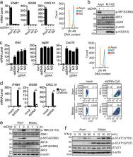
          The cyclic GMP-AMP synthase (cGAS) is a widely used DNA sensor, which detects cytosolic DNA species without a preference of self or non-self microbial DNA in interphase to initiate innate immune response. How cGAS is regulated to avoid self-DNA sensing upon nuclear envelope breakdown (NEBD) during mitosis remains enigmatic. Here we show that cGAS is mostly localized in the cytoplasm in interphase and rapidly translocated to chromosomes upon NEBD in mitosis. The major mitotic kinase CDK1-cyclin B complex phosphorylates human cGAS at S305 or mouse cGAS at S291, which inhibits its ability to synthesize cGAMP upon mitotic entry. The type 1 phosphatase PP1 dephosphorylates cGAS upon mitotic exit to enable its DNA sensing ability. Our findings reveal a mechanism on how the DNA sensor cGAS is post-translationally regulated by cell cycle-dependent enzymes to ensure its proper activation for host defense of cytosolic DNA in interphase and inert to self-DNA in mitosis.

          Related collections

          Most cited references21

          • Record: found
          • Abstract: found
          • Article: not found

          DNA damage primes the type I interferon system via the cytosolic DNA sensor STING to promote anti-microbial innate immunity.

          Dysfunction in Ataxia-telangiectasia mutated (ATM), a central component of the DNA repair machinery, results in Ataxia Telangiectasia (AT), a cancer-prone disease with a variety of inflammatory manifestations. By analyzing AT patient samples and Atm(-/-) mice, we found that unrepaired DNA lesions induce type I interferons (IFNs), resulting in enhanced anti-viral and anti-bacterial responses in Atm(-/-) mice. Priming of the type I interferon system by DNA damage involved release of DNA into the cytoplasm where it activated the cytosolic DNA sensing STING-mediated pathway, which in turn enhanced responses to innate stimuli by activating the expression of Toll-like receptors, RIG-I-like receptors, cytoplasmic DNA sensors, and their downstream signaling partners. This study provides a potential explanation for the inflammatory phenotype of AT patients and establishes damaged DNA as a cell intrinsic danger signal that primes the innate immune system for a rapid and amplified response to microbial and environmental threats.
            Bookmark
            • Record: found
            • Abstract: found
            • Article: not found

            Sumoylation Promotes the Stability of the DNA Sensor cGAS and the Adaptor STING to Regulate the Kinetics of Response to DNA Virus.

            During viral infection, sensing of cytosolic DNA by the cyclic GMP-AMP synthase (cGAS) activates the adaptor protein STING and triggers an antiviral response. Little is known about the mechanisms that determine the kinetics of activation and deactivation of the cGAS-STING pathway, ensuring effective but controlled innate antiviral responses. Here we found that the ubiquitin ligase Trim38 targets cGas for sumoylation in uninfected cells and during the early phase of viral infection. Sumoylation of cGas prevented its polyubiquitination and degradation. Trim38 also sumoylated Sting during the early phase of viral infection, promoting both Sting activation and protein stability. In the late phase of infection, cGas and Sting were desumoylated by Senp2 and subsequently degraded via proteasomal and chaperone-mediated autophagy pathways, respectively. Our findings reveal an essential role for Trim38 in the innate immune response to DNA virus and provide insight into the mechanisms that ensure optimal activation and deactivation of the cGAS-STING pathway.
              Bookmark
              • Record: found
              • Abstract: found
              • Article: not found

              A Circular RNA Protects Dormant Hematopoietic Stem Cells from DNA Sensor cGAS-Mediated Exhaustion

              Disrupting the balance between self-renewal and differentiation of hematopoietic stem cells (HSCs) leads to bone marrow failure or hematologic malignancy. However, how HSCs sustain their quiescent state and avoid type I interferon (IFN)-mediated exhaustion remains elusive. Here we defined a circular RNA that we named cia-cGAS that was highly expressed in the nucleus of long-term (LT)-HSCs. Cia-cGAS deficiency in mice caused elevated expression of type I IFNs in bone marrow and led to decreased numbers of dormant LT-HSCs. Under homeostatic conditions, cia-cGAS bound DNA sensor cGAS in the nucleus to block its synthase activity, thereby protecting dormant LT-HSCs from cGAS-mediated exhaustion. Moreover, cia-cGAS harbored a stronger binding affinity to cGAS than self-DNA did and consequently suppressed cGAS-mediated production of type I IFNs in LT-HSCs. Our findings reveal a mechanism by which cia-cGAS inhibits nuclear cGAS by blocking its enzymatic activity and preventing cGAS from recognizing self-DNA to maintain host homeostasis.
                Bookmark

                Author and article information

                Contributors
                shuh@whu.edu.cn
                Journal
                Cell Discov
                Cell Discov
                Cell Discovery
                Springer Singapore (Singapore )
                2056-5968
                28 April 2020
                28 April 2020
                2020
                : 6
                : 26
                Affiliations
                ISNI 0000 0001 2331 6153, GRID grid.49470.3e, Department of Infectious Diseases, Zhongnan Hospital of Wuhan University, Frontier Science Center for Immunology and Metabolism Medical Research Institute, , Wuhan University, ; Wuhan, 430071 China
                Author information
                http://orcid.org/0000-0003-3094-4806
                http://orcid.org/0000-0001-9102-3272
                Article
                162
                10.1038/s41421-020-0162-2
                7186227
                32351706
                acc99453-9221-443f-b487-9e258fcbc1a6
                © The Author(s) 2020

                Open Access This article is licensed under a Creative Commons Attribution 4.0 International License, which permits use, sharing, adaptation, distribution and reproduction in any medium or format, as long as you give appropriate credit to the original author(s) and the source, provide a link to the Creative Commons license, and indicate if changes were made. The images or other third party material in this article are included in the article’s Creative Commons license, unless indicated otherwise in a credit line to the material. If material is not included in the article’s Creative Commons license and your intended use is not permitted by statutory regulation or exceeds the permitted use, you will need to obtain permission directly from the copyright holder. To view a copy of this license, visit http://creativecommons.org/licenses/by/4.0/.

                History
                : 15 January 2020
                : 12 March 2020
                Funding
                Funded by: FundRef https://doi.org/10.13039/501100001809, National Natural Science Foundation of China (National Science Foundation of China);
                Award ID: 31830024
                Award ID: 31630045
                Award Recipient :
                Funded by: This work was supported by grants from the State Key R&D Program of China (2017YFA0505800, 2016YFA0502102), and the National Natural Science Foundation of China (31830024, 31630045).
                Categories
                Article
                Custom metadata
                © The Author(s) 2020

                innate immunity,cell division
                innate immunity, cell division

                Comments

                Comment on this article