- Record: found
- Abstract: found
- Article: found
Neurovascular coupling and oxygenation are decreased in hippocampus compared to neocortex because of microvascular differences

Read this article at
Abstract
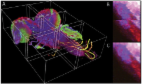
The hippocampus is essential for spatial and episodic memory but is damaged early in Alzheimer’s disease and is very sensitive to hypoxia. Understanding how it regulates its oxygen supply is therefore key for designing interventions to preserve its function. However, studies of neurovascular function in the hippocampus in vivo have been limited by its relative inaccessibility. Here we compared hippocampal and visual cortical neurovascular function in awake mice, using two photon imaging of individual neurons and vessels and measures of regional blood flow and haemoglobin oxygenation. We show that blood flow, blood oxygenation and neurovascular coupling were decreased in the hippocampus compared to neocortex, because of differences in both the vascular network and pericyte and endothelial cell function. Modelling oxygen diffusion indicates that these features of the hippocampal vasculature may restrict oxygen availability and could explain its sensitivity to damage during neurological conditions, including Alzheimer’s disease, where the brain’s energy supply is decreased.
Abstract
The hippocampus is particularly sensitive to hypoxia but it has been difficult to study blood flow in this region. Here the authors compare the neurovascular function of the hippocampus and cortex and in awake mice, and find differences associated with microvascular structure.
Related collections
Most cited references67
- Record: found
- Abstract: found
- Article: found
Molecular Architecture of the Mouse Nervous System
- Record: found
- Abstract: found
- Article: not found
Blood-brain barrier breakdown in the aging human hippocampus.
- Record: found
- Abstract: found
- Article: found
Globally optimal stitching of tiled 3D microscopic image acquisitions
Author and article information
Comments
Comment on this article
See how this article has been cited at scite.ai
scite shows how a scientific paper has been cited by providing the context of the citation, a classification describing whether it supports, mentions, or contrasts the cited claim, and a label indicating in which section the citation was made.