1
views
0
recommends
+1 Recommend
0 collections
    0
    shares
      • Record: found
      • Abstract: found
      • Article: found
      Is Open Access

      Fast three‐dimensional image generation for healthy brain aging using diffeomorphic registration

      other

      Read this article at

      Bookmark
          There is no author summary for this article yet. Authors can add summaries to their articles on ScienceOpen to make them more accessible to a non-specialist audience.

          Abstract

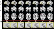
          Predicting brain aging can help in the early detection and prognosis of neurodegenerative diseases. Longitudinal cohorts of healthy subjects scanned through magnetic resonance imaging (MRI) have been essential to understand the structural brain changes due to aging. However, these cohorts suffer from missing data due to logistic issues in the recruitment of subjects. This paper proposes a methodology for filling up missing data in longitudinal cohorts with anatomically plausible images that capture the subject‐specific aging process. The proposed methodology is developed within the framework of diffeomorphic registration. First, two novel modules are introduced within Synthmorph, a fast, state‐of‐the‐art deep learning‐based diffeomorphic registration method, to simulate the aging process between the first and last available MRI scan for each subject in three‐dimensional (3D). The use of image registration also makes the generated images plausible by construction. Second, we used six image similarity measurements to rearrange the generated images to the specific age range. Finally, we estimated the age of every generated image by using the assumption of linear brain decay in healthy subjects. The methodology was evaluated on 2662 T1‐weighted MRI scans from 796 healthy participants from 3 different longitudinal cohorts: Alzheimer's Disease Neuroimaging Initiative, Open Access Series of Imaging Studies‐3, and Group of Neuropsychological Studies of the Canary Islands (GENIC). In total, we generated 7548 images to simulate the access of a scan per subject every 6 months in these cohorts. We evaluated the quality of the synthetic images using six quantitative measurements and a qualitative assessment by an experienced neuroradiologist with state‐of‐the‐art results. The assumption of linear brain decay was accurate in these cohorts ( R 2 ∈ [.924, .940]). The experimental results show that the proposed methodology can produce anatomically plausible aging predictions that can be used to enhance longitudinal datasets. Compared to deep learning‐based generative methods, diffeomorphic registration is more likely to preserve the anatomy of the different structures of the brain, which makes it more appropriate for its use in clinical applications. The proposed methodology is able to efficiently simulate anatomically plausible 3D MRI scans of brain aging of healthy subjects from two images scanned at two different time points.

          Abstract

          In this work, we proposed a methodology with the aim of simulating subject‐specific aging in brain magnetic resonance imaging (MRI) given two three‐dimensional images acquired at different time points. Deep learning‐based diffeomorphic registration was used as a backbone to generate deformation fields at different integration points. Similarity measurements were used for controlling the age estimation of the generated images by using a linear assumption.

          Related collections

          Most cited references73

          • Record: found
          • Abstract: not found
          • Book Chapter: not found

          U-Net: Convolutional Networks for Biomedical Image Segmentation

            Bookmark
            • Record: found
            • Abstract: found
            • Article: not found

            FreeSurfer.

            FreeSurfer is a suite of tools for the analysis of neuroimaging data that provides an array of algorithms to quantify the functional, connectional and structural properties of the human brain. It has evolved from a package primarily aimed at generating surface representations of the cerebral cortex into one that automatically creates models of most macroscopically visible structures in the human brain given any reasonable T1-weighted input image. It is freely available, runs on a wide variety of hardware and software platforms, and is open source. Copyright © 2012 Elsevier Inc. All rights reserved.
              Bookmark
              • Record: found
              • Abstract: found
              • Article: not found

              A fast diffeomorphic image registration algorithm.

              This paper describes DARTEL, which is an algorithm for diffeomorphic image registration. It is implemented for both 2D and 3D image registration and has been formulated to include an option for estimating inverse consistent deformations. Nonlinear registration is considered as a local optimisation problem, which is solved using a Levenberg-Marquardt strategy. The necessary matrix solutions are obtained in reasonable time using a multigrid method. A constant Eulerian velocity framework is used, which allows a rapid scaling and squaring method to be used in the computations. DARTEL has been applied to intersubject registration of 471 whole brain images, and the resulting deformations were evaluated in terms of how well they encode the shape information necessary to separate male and female subjects and to predict the ages of the subjects.
                Bookmark

                Author and article information

                Contributors
                jingruf@kth.se
                Journal
                Hum Brain Mapp
                Hum Brain Mapp
                10.1002/(ISSN)1097-0193
                HBM
                Human Brain Mapping
                John Wiley & Sons, Inc. (Hoboken, USA )
                1065-9471
                1097-0193
                05 December 2022
                March 2023
                : 44
                : 4 ( doiID: 10.1002/hbm.v44.4 )
                : 1289-1308
                Affiliations
                [ 1 ] Division of Biomedical Imaging Department of Biomedical Engineering and Health Systems, KTH Royal Institute of Technology Stockholm Sweden
                [ 2 ] Division of Radiology Department for Clinical Science, Intervention and Technology (CLINTEC), Karolinska Institutet Stockholm Sweden
                [ 3 ] Medical Radiation Physics and Nuclear Medicine Functional Unit of Nuclear Medicine, Karolinska University Hospital Huddinge Stockholm Sweden
                [ 4 ] Department of Psychology Faculty of Health Sciences, University Fernando Pessoa Canarias Las Palmas Spain
                [ 5 ] Division of Clinical Geriatrics Centre for Alzheimer Research, Department of Neurobiology, Care Sciences, and Society (NVS), Karolinska Institutet Stockholm Sweden
                [ 6 ] Department of Neuroimaging Centre for Neuroimaging Sciences, Institute of Psychiatry, Psychology and Neuroscience, King's College London London United Kingdom
                Author notes
                [*] [* ] Correspondence

                Jingru Fu, Division of Biomedical Imaging, KTH Royal Institute of Technology, Stockholm, Sweden.

                Email: jingruf@ 123456kth.se

                Author information
                https://orcid.org/0000-0003-4175-395X
                https://orcid.org/0000-0001-7563-732X
                https://orcid.org/0000-0002-3115-2977
                https://orcid.org/0000-0001-9522-4338
                https://orcid.org/0000-0001-5765-2964
                Article
                HBM26165
                10.1002/hbm.26165
                9921328
                36468536
                ab19ef65-ace7-44a0-986c-a7ad0d9515f8
                © 2022 The Authors. Human Brain Mapping published by Wiley Periodicals LLC.

                This is an open access article under the terms of the http://creativecommons.org/licenses/by-nc-nd/4.0/ License, which permits use and distribution in any medium, provided the original work is properly cited, the use is non‐commercial and no modifications or adaptations are made.

                History
                : 15 November 2022
                : 16 June 2022
                : 16 November 2022
                Page count
                Figures: 12, Tables: 8, Pages: 20, Words: 15365
                Funding
                Funded by: Digital Futures
                Award ID: project dBrain
                Funded by: VINNOVA , doi 10.13039/501100001858;
                Award ID: 2108
                Funded by: Alzheimer's Disease Neuroimaging Initiative , doi 10.13039/100007333;
                Funded by: National Institutes of Health , doi 10.13039/100000002;
                Award ID: U01 AG024904
                Funded by: DOD ADNI
                Award ID: W81XWH‐12‐2‐0012
                Funded by: National Institute on Aging , doi 10.13039/100000049;
                Funded by: National Institute of Biomedical Imaging and Bioengineering , doi 10.13039/100000070;
                Funded by: AbbVie , doi 10.13039/100006483;
                Funded by: Alzheimer's Association , doi 10.13039/100000957;
                Funded by: Alzheimer's Drug Discovery Foundation , doi 10.13039/100002565;
                Funded by: Araclon Biotech
                Funded by: BioClinica, Inc. , doi 10.13039/100007742;
                Funded by: Biogen , doi 10.13039/100005614;
                Funded by: Bristol‐Myers Squibb
                Funded by: CereSpir, Inc.
                Funded by: Cogstate
                Funded by: Eisai Inc. , doi 10.13039/501100004896;
                Funded by: Elan Pharmaceuticals, Inc.
                Funded by: Eli Lilly and Company , doi 10.13039/100004312;
                Funded by: EUROIMMUN Medizinische Labordiagnostika AG
                Funded by: F. Hoffmann‐La Roche Ltd
                Funded by: Genentech, Inc. , doi 10.13039/100004328;
                Funded by: Fujirebio US , doi 10.13039/100014400;
                Funded by: GE Healthcare , doi 10.13039/100006775;
                Funded by: IXICO Ltd , doi 10.13039/501100015725;
                Funded by: Janssen Alzheimer Immunotherapy Research & Development, LLC
                Funded by: Johnson & Johnson Pharmaceutical Research & Development LLC
                Funded by: Lumosity
                Funded by: Lundbeck , doi 10.13039/501100013327;
                Funded by: Merck & Co., Inc. , doi 10.13039/100004334;
                Funded by: Meso Scale Diagnostics, LLC. , doi 10.13039/100007054;
                Funded by: NeuroRx Research
                Funded by: Neurotrack Technologies
                Funded by: Novartis Pharmaceuticals Corporation , doi 10.13039/100008272;
                Funded by: Pfizer Inc. , doi 10.13039/100004319;
                Funded by: Piramal Imaging
                Funded by: Servier , doi 10.13039/501100011725;
                Funded by: Takeda Pharmaceutical Company , doi 10.13039/100007723;
                Funded by: Transition Therapeutics
                Funded by: Canadian Institutes of Health Research , doi 10.13039/501100000024;
                Funded by: China Scholarship Council , doi 10.13039/501100004543;
                Funded by: Center for Innovative Medicine (CIMED) , doi 10.13039/501100018713;
                Funded by: Stockholm County Council and Karolinska Institutet
                Funded by: Hjärnfonden , doi 10.13039/501100003792;
                Funded by: Alzheimerfonden , doi 10.13039/501100008599;
                Funded by: Demensfonden , doi 10.13039/501100021594;
                Funded by: Neurofonden
                Funded by: Stiftelsen För Gamla Tjänarinnor , doi 10.13039/100010815;
                Funded by: Fundación Canaria Dr Manuel Morales
                Funded by: Fundación Cajacanarias , doi 10.13039/100012000;
                Funded by: Estrategia de Especialización Inteligente de Canarias RIS3 from Consejería de Economía
                Funded by: Industria, Comercio y Conocimiento del Gobierno de Canarias
                Funded by: Programa Operativo FEDER Canarias 2014–2020
                Award ID: ProID2020010063
                Funded by: Barncancerfonden , doi 10.13039/501100006313;
                Award ID: MT2019‐0019
                Categories
                Technical Report
                Technical Report
                Custom metadata
                2.0
                March 2023
                Converter:WILEY_ML3GV2_TO_JATSPMC version:6.2.5 mode:remove_FC converted:11.02.2023

                Neurology
                brain aging,diffeomorphic registration,medical image generation,synthetic brain aging

                Comments

                Comment on this article