14
views
0
recommends
+1 Recommend
0 collections
    0
    shares
      • Record: found
      • Abstract: found
      • Article: found
      Is Open Access

      Atypical Integration of Sensory-to-Transmodal Functional Systems Mediates Symptom Severity in Autism

      research-article

      Read this article at

      Bookmark
          There is no author summary for this article yet. Authors can add summaries to their articles on ScienceOpen to make them more accessible to a non-specialist audience.

          Abstract

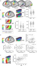
          A notable characteristic of autism spectrum disorder (ASD) is co-occurring deficits in low-level sensory processing and high-order social interaction. While there is evidence indicating detrimental cascading effects of sensory anomalies on the high-order cognitive functions in ASD, the exact pathological mechanism underlying their atypical functional interaction across the cortical hierarchy has not been systematically investigated. To address this gap, here we assessed the functional organisation of sensory and motor areas in ASD, and their relationship with subcortical and high-order trandmodal systems. In a resting-state fMRI data of 107 ASD and 113 neurotypical individuals, we applied advanced connectopic mapping to probe functional organization of primary sensory/motor areas, together with targeted seed-based intrinsic functional connectivity (iFC) analyses. In ASD, the connectopic mapping revealed topological anomalies (i.e., excessively more segregated iFC) in the motor and visual areas, the former of which patterns showed association with the symptom severity of restricted and repetitive behaviors. Moreover, the seed-based analysis found diverging patterns of ASD-related connectopathies: decreased iFCs within the sensory/motor areas but increased iFCs between sensory and subcortical structures. While decreased iFCs were also found within the higher-order functional systems, the overall proportion of this anomaly tends to increase along the level of cortical hierarchy, suggesting more dysconnectivity in the higher-order functional networks. Finally, we demonstrated that the association between low-level sensory/motor iFCs and clinical symptoms in ASD was mediated by the high-order transmodal systems, suggesting pathogenic functional interactions along the cortical hierarchy. Findings were largely replicated in the independent dataset. These results highlight that atypical integration of sensory-to-high-order systems contributes to the complex ASD symptomatology.

          Related collections

          Most cited references114

          • Record: found
          • Abstract: found
          • Article: not found

          The organization of the human cerebral cortex estimated by intrinsic functional connectivity.

          Information processing in the cerebral cortex involves interactions among distributed areas. Anatomical connectivity suggests that certain areas form local hierarchical relations such as within the visual system. Other connectivity patterns, particularly among association areas, suggest the presence of large-scale circuits without clear hierarchical relations. In this study the organization of networks in the human cerebrum was explored using resting-state functional connectivity MRI. Data from 1,000 subjects were registered using surface-based alignment. A clustering approach was employed to identify and replicate networks of functionally coupled regions across the cerebral cortex. The results revealed local networks confined to sensory and motor cortices as well as distributed networks of association regions. Within the sensory and motor cortices, functional connectivity followed topographic representations across adjacent areas. In association cortex, the connectivity patterns often showed abrupt transitions between network boundaries. Focused analyses were performed to better understand properties of network connectivity. A canonical sensory-motor pathway involving primary visual area, putative middle temporal area complex (MT+), lateral intraparietal area, and frontal eye field was analyzed to explore how interactions might arise within and between networks. Results showed that adjacent regions of the MT+ complex demonstrate differential connectivity consistent with a hierarchical pathway that spans networks. The functional connectivity of parietal and prefrontal association cortices was next explored. Distinct connectivity profiles of neighboring regions suggest they participate in distributed networks that, while showing evidence for interactions, are embedded within largely parallel, interdigitated circuits. We conclude by discussing the organization of these large-scale cerebral networks in relation to monkey anatomy and their potential evolutionary expansion in humans to support cognition.
            Bookmark
            • Record: found
            • Abstract: found
            • Article: not found

            The minimal preprocessing pipelines for the Human Connectome Project.

            The Human Connectome Project (HCP) faces the challenging task of bringing multiple magnetic resonance imaging (MRI) modalities together in a common automated preprocessing framework across a large cohort of subjects. The MRI data acquired by the HCP differ in many ways from data acquired on conventional 3 Tesla scanners and often require newly developed preprocessing methods. We describe the minimal preprocessing pipelines for structural, functional, and diffusion MRI that were developed by the HCP to accomplish many low level tasks, including spatial artifact/distortion removal, surface generation, cross-modal registration, and alignment to standard space. These pipelines are specially designed to capitalize on the high quality data offered by the HCP. The final standard space makes use of a recently introduced CIFTI file format and the associated grayordinate spatial coordinate system. This allows for combined cortical surface and subcortical volume analyses while reducing the storage and processing requirements for high spatial and temporal resolution data. Here, we provide the minimum image acquisition requirements for the HCP minimal preprocessing pipelines and additional advice for investigators interested in replicating the HCP's acquisition protocols or using these pipelines. Finally, we discuss some potential future improvements to the pipelines. Copyright © 2013 Elsevier Inc. All rights reserved.
              Bookmark
              • Record: found
              • Abstract: found
              • Article: not found

              Whole brain segmentation: automated labeling of neuroanatomical structures in the human brain.

              We present a technique for automatically assigning a neuroanatomical label to each voxel in an MRI volume based on probabilistic information automatically estimated from a manually labeled training set. In contrast to existing segmentation procedures that only label a small number of tissue classes, the current method assigns one of 37 labels to each voxel, including left and right caudate, putamen, pallidum, thalamus, lateral ventricles, hippocampus, and amygdala. The classification technique employs a registration procedure that is robust to anatomical variability, including the ventricular enlargement typically associated with neurological diseases and aging. The technique is shown to be comparable in accuracy to manual labeling, and of sufficient sensitivity to robustly detect changes in the volume of noncortical structures that presage the onset of probable Alzheimer's disease.
                Bookmark

                Author and article information

                Contributors
                Journal
                Front Psychiatry
                Front Psychiatry
                Front. Psychiatry
                Frontiers in Psychiatry
                Frontiers Media S.A.
                1664-0640
                20 August 2021
                2021
                : 12
                : 699813
                Affiliations
                [1] 1Institute for Basic Science, Center for Neuroscience Imaging Research, Sungkyunkwan University , Suwon, South Korea
                [2] 2Department of Biomedical Engineering, Sungkyunkwan University , Suwon, South Korea
                [3] 3Donders Institute of Brain, Cognition, and Behaviour, Radboud University Medical Center , Nijmegen, Netherlands
                [4] 4Otto Hahn Group Cognitive Neurogenetics, Max Planck Institute for Human Cognitive and Brain Sciences , Leipzig, Germany
                [5] 5Institute of Neuroscience and Medicine (INM-7), Forschungszentrum Jülich , Jülich, Germany
                [6] 6Department of Psychiatry, Autism Research Centre, University of Cambridge , Cambridge, United Kingdom
                [7] 7Brain Mapping Unit, Department of Psychiatry, University of Cambridge , Cambridge, United Kingdom
                [8] 8Center for the Developing Brain, Child Mind Institute , New York, NY, United States
                [9] 9Center for Biomedical Imaging and Neuromodulation, Nathan Kline Institute , New York, NY, United States
                [10] 10McConnell Brain Imaging Centre, Montreal Neurological Institute and Hospital, McGill University , Montreal, QC, Canada
                [11] 11Autism Center, Child Mind Institute , New York, NY, United States
                Author notes

                Edited by: Ryu-ichiro Hashimoto, Showa University, Japan

                Reviewed by: Deniz Vatansever, Fudan University, China; Matthew W. Mosconi, University of Kansas, United States

                *Correspondence: Seok-Jun Hong hong.seok.jun@ 123456gmail.com

                This article was submitted to Computational Psychiatry, a section of the journal Frontiers in Psychiatry

                Article
                10.3389/fpsyt.2021.699813
                8417581
                34489757
                aaed26ba-edf0-465e-8468-b6c31de27f7b
                Copyright © 2021 Park, Haak, Cho, Valk, Bethlehem, Milham, Bernhardt, Di Martino and Hong.

                This is an open-access article distributed under the terms of the Creative Commons Attribution License (CC BY). The use, distribution or reproduction in other forums is permitted, provided the original author(s) and the copyright owner(s) are credited and that the original publication in this journal is cited, in accordance with accepted academic practice. No use, distribution or reproduction is permitted which does not comply with these terms.

                History
                : 24 April 2021
                : 16 July 2021
                Page count
                Figures: 5, Tables: 1, Equations: 0, References: 114, Pages: 16, Words: 10811
                Categories
                Psychiatry
                Original Research

                Clinical Psychology & Psychiatry
                autism spectrum disoder,cortical hierarchy,low-level sensory,high-order system,connectopic mapping,subcortico-cortical connectivity

                Comments

                Comment on this article