3
views
0
recommends
+1 Recommend
1 collections
    0
    shares

         An official journal of the Society for Reproduction and Fertility. Learn more

      • Record: found
      • Abstract: found
      • Article: found
      Is Open Access

      Incidence of pregnancy loss and characterization of fetal development in red pandas

      research-article

      Read this article at

      ScienceOpenPublisherPMC
      Bookmark
          There is no author summary for this article yet. Authors can add summaries to their articles on ScienceOpen to make them more accessible to a non-specialist audience.

          Abstract

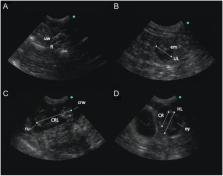
          Previous reports indicate that red pandas ( Ailurus fulgens styani) may experience fetal loss during gestation; however, neither the rate nor timing of pregnancy failure has been described in this species. The objective of this study was to utilize ultrasound video and images collected between 2010 and 2020 at the Cincinnati Zoo and Botanical Garden to better characterize pregnancy loss and fetal development. Trans-abdominal ultrasound examinations were performed on six female red pandas over a 10-year period, resulting in 12 profiles. Pregnancy was diagnosed via ultrasound in 10 of 12 profiles, and 40.0% of pregnancies showed evidence of fetal loss prior to parturition. Pregnancy loss was classified into lost (2 of 10; 20.0%), in which no cubs were produced, or partial loss (2 of 10; 20.0%), in which two concepti were visualized via ultrasound, but only one cub was born. Fetal loss occurred between days 51 and 23 pre-partum. Fetal growth characteristics were documented, including skeletal ossification (occurring between days 32 and 27 pre-partum), crown-rump length, head length, cranial length, and fetal heart rate (173–206 b.p.m.). These findings provide novel insights into pregnancy loss, may serve as a reference for milestones of fetal development, and may be useful in diagnosing pregnancy and assessing pregnancy loss in red pandas.

          Lay summary

          For many wildlife species, there is no non-invasive method of determining pregnancy; therefore, the rate of pregnancy loss oftentimes is unknown. Many red pandas in human care that are paired for breeding are observed exhibiting normal mating behaviors; however, only a relatively low proportion of females produce cubs. We utilized animals conditioned for ultrasound examination to diagnose pregnancy and characterize the incidence and timing of pregnancy loss. In total, 12 potential pregnancies were monitored, beginning after breeding season and ending ~2 weeks prior to anticipated cubbing. Of these, ten were (83.3%) were diagnosed as pregnant, with 40% undergoing either full or partial pregnancy loss. Fetal growth characteristics, such as body length and head size, are described which may be useful for monitoring pregnancies and estimating fetal age. Results of this study provide novel data on pregnancy loss in red pandas. Insights into the rate and timing of reproductive failure may illuminate causes and contributing factors, ultimately allowing for improvements in husbandry which may result in greater reproductive success of individuals recommended for breeding.

          Related collections

          Most cited references24

          • Record: found
          • Abstract: found
          • Article: found
          Is Open Access

          Genomic evidence for two phylogenetic species and long-term population bottlenecks in red pandas

          Population genomics reveal two species in red pandas, correct their species boundaries, and reconstruct their evolutionary histories.
            Bookmark
            • Record: found
            • Abstract: found
            • Article: not found

            Canine embryonic and fetal development: a review.

            S Pretzer (2008)
            The progression from a fertilized oocyte to a newborn puppy is a remarkable phenomenon that occurs in a period of approximately two months. Embryonic development encompasses the period of time at which three germ layers differentiate: ectoderm, mesoderm, and endoderm. Organ systems are formed from these germ layers, with most of the reproductive tract being derived from mesoderm. Organogenesis is complete prior to the fetal stage in canine embryos, but sexual differentiation occurs during the fetal stage. Sexual differentiation is a well-coordinated progression of events that is directed initially by the genotype of the developing embryo and fetus. Developing fetuses are inherently female and will develop as such in the absence of a Y chromosome. Male fetuses develop as the Y chromosome causes regression of the female duct system and development of the male duct system. Testicular descent in the canine begins in the fetal stage, but is not completed until after birth.
              Bookmark
              • Record: found
              • Abstract: found
              • Article: found
              Is Open Access

              Early detection and staging of spontaneous embryo resorption by ultrasound biomicroscopy in murine pregnancy

              Background Embryo resorption is a major problem in human medicine, agricultural animal production and in conservation breeding programs. Underlying mechanisms have been investigated in the well characterised mouse model. However, post mortem studies are limited by the rapid disintegration of embryonic structures. A method to reliably identify embryo resorption in alive animals has not been established yet. In our study we aim to detect embryos undergoing resorption in vivo at the earliest possible stage by ultra-high frequency ultrasound. Methods In a longitudinal study, we monitored 30 pregnancies of wild type C57BI/6 mice using ultra-high frequency ultrasound (30-70 MHz), so called ultrasound biomicroscopy (UBM). We compared the sonoembryology of mouse conceptuses under spontaneous resorption and neighbouring healthy conceptuses and correlated the live ultrasound data with the respective histology. Results The process of embryo resorption comprised of four stages: first, the conceptus exhibited growth retardation, second, bradycardia and pericardial edema were observed, third, further development ceased and the embryo died, and finally embryo remnants were resorbed by maternal immune cells. In early gestation (day 7 and 8), growth retardation was characterized by a small embryonic cavity. The embryo and its membranes were ill defined or did not develop at all. The echodensity of the embryonic fluid increased and within one to two days, the embryo and its cavity disappeared and was transformed into echodense tissue surrounded by fluid filled caverns. In corresponding histologic preparations, fibrinoid material interspersed with maternal granulocytes and lacunae filled with maternal blood were observed. In later stages (day 9–11) resorption prone embryos were one day behind in their development compared to their normal siblings. The space between Reichert’s membrane and inner yolk sac membrane was enlarged The growth retarded embryos exhibited bradycardia and ultimately cessation of heart beat. Corresponding histology showed apoptotic cells in the embryo while the placenta was still intact. In the subsequent resorption process first the embryo and then its membranes disappeared. Conclusions Our results provide a temporal time course of embryo resorption. With this method, animals exhibiting embryo resorption can be targeted, enabling the investigation of underlying mechanisms before the onset of total embryo disintegration.
                Bookmark

                Author and article information

                Journal
                Reprod Fertil
                Reprod Fertil
                raf
                Reproduction & Fertility
                Bioscientifica Ltd (Bristol )
                2633-8386
                December 2021
                21 October 2021
                : 2
                : 4
                : 292-299
                Affiliations
                [1 ]Center for Conservation and Research of Endangered Wildlife , Cincinnati Zoo & Botanical Garden, Cincinnati, Ohio, USA
                Author notes
                Correspondence should be addressed to E Curry: erin.curry@ 123456cincinnatizoo.org

                *(J Lowe is currently at Meinig School of Biomedical Engineering, Cornell University, Ithaca, New York, USA)

                Article
                RAF-21-0079
                10.1530/RAF-21-0079
                8801018
                35118406
                aab9ef5f-f3df-41f1-8680-a1e75c16010b
                © The authors

                This work is licensed under a Creative Commons Attribution 4.0 International License.

                History
                : 08 October 2021
                : 21 October 2021
                Categories
                Research

                carnivore reproduction,delayed implantation,embryonic diapause,wildlife,pseudopregnancy,ultrasonography

                Comments

                Comment on this article