1
views
0
recommends
+1 Recommend
0 collections
    0
    shares
      • Record: found
      • Abstract: found
      • Article: found
      Is Open Access

      Spatial analysis of histology in 3D: quantification and visualization of organ and tumor level tissue environment

      research-article

      Read this article at

      Bookmark
          There is no author summary for this article yet. Authors can add summaries to their articles on ScienceOpen to make them more accessible to a non-specialist audience.

          Abstract

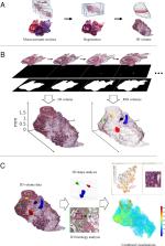
          Histological changes in tissue are of primary importance in pathological research and diagnosis. Automated histological analysis requires ability to computationally separate pathological alterations from normal tissue. Conventional histopathological assessments are performed from individual tissue sections, leading to the loss of three-dimensional context of the tissue. Yet, the tissue context and spatial determinants are critical in several pathologies, such as in understanding growth patterns of cancer in its local environment. Here, we develop computational methods for visualization and quantitative assessment of histopathological alterations in three dimensions. First, we reconstruct the 3D representation of the whole organ from serial sectioned tissue. Then, we proceed to analyze the histological characteristics and regions of interest in 3D. As our example cases, we use whole slide images representing hematoxylin-eosin stained whole mouse prostates in a Pten+/- mouse prostate tumor model. We show that quantitative assessment of tumor sizes, shapes, and separation between spatial locations within the organ enable characterizing and grouping tumors. Further, we show that 3D visualization of tissue with computationally quantified features provides an intuitive way to observe tissue pathology. Our results underline the heterogeneity in composition and cellular organization within individual tumors. As an example, we show how prostate tumors have nuclear density gradients indicating areas of tumor growth directions and reflecting varying pressure from the surrounding tissue. The methods presented here are applicable to any tissue and different types of pathologies. This work provides a proof-of-principle for gaining a comprehensive view from histology by studying it quantitatively in 3D.

          Abstract

          Histology; 3D reconstruction; Quantitative imaging; Tissue analysis; Spatial analysis; Image analysis; Visualization

          Related collections

          Most cited references42

          • Record: found
          • Abstract: found
          • Article: not found

          Fiji: an open-source platform for biological-image analysis.

          Fiji is a distribution of the popular open-source software ImageJ focused on biological-image analysis. Fiji uses modern software engineering practices to combine powerful software libraries with a broad range of scripting languages to enable rapid prototyping of image-processing algorithms. Fiji facilitates the transformation of new algorithms into ImageJ plugins that can be shared with end users through an integrated update system. We propose Fiji as a platform for productive collaboration between computer science and biology research communities.
            Bookmark
            • Record: found
            • Abstract: not found
            • Article: not found

            A Threshold Selection Method from Gray-Level Histograms

              Bookmark
              • Record: found
              • Abstract: found
              • Article: found
              Is Open Access

              Animal models of necrotizing enterocolitis: review of the literature and state of the art

              Abstract Necrotizing enterocolitis (NEC) remains the leading cause of gastrointestinal surgical emergency in preterm neonates. Over the last five decades, a variety of experimental models have been developed to study the pathophysiology of this disease and to test the effectiveness of novel therapeutic strategies. Experimental NEC is mainly modeled in neonatal rats, mice and piglets. In this review, we focus on these experimental models and discuss the major advantages and disadvantages of each. We also briefly discuss other models that are not as widely used but have contributed to our current knowledge of NEC.
                Bookmark

                Author and article information

                Contributors
                Journal
                Heliyon
                Heliyon
                Heliyon
                Elsevier
                2405-8440
                14 January 2022
                January 2022
                14 January 2022
                : 8
                : 1
                : e08762
                Affiliations
                [a ]Institute of Biomedicine, University of Turku, Turku, Finland
                [b ]Faculty of Medicine and Health Technology, Tampere University, Finland
                [c ]Tays Cancer Center, Tampere University Hospital, Tampere, Finland
                [d ]Fimlab Laboratories Ltd, Tampere University Hospital, Tampere, Finland
                [e ]Institute of Biomedicine, University of Eastern Finland, Kuopio, Finland
                Author notes
                [* ]Corresponding author at: Institute of Biomedicine, University of Turku, Turku, Finland. pekka.ruusuvuori@ 123456utu.fi
                [1]

                These authors contributed equally.

                Article
                S2405-8440(22)00050-0 e08762
                10.1016/j.heliyon.2022.e08762
                8800033
                aa815401-b4ab-4368-b071-557dde5b45df
                © 2022 The Authors

                This is an open access article under the CC BY-NC-ND license (http://creativecommons.org/licenses/by-nc-nd/4.0/).

                History
                : 27 October 2021
                : 24 November 2021
                : 11 January 2022
                Categories
                Research Article

                histology,3d reconstruction,quantitative imaging,tissue analysis,spatial analysis,image analysis,visualization

                Comments

                Comment on this article