46
views
0
recommends
+1 Recommend
0 collections
    0
    shares
      • Record: found
      • Abstract: found
      • Article: not found

      The Rab5 Effector Rabankyrin-5 Regulates and Coordinates Different Endocytic Mechanisms

      research-article

      Read this article at

      ScienceOpenPublisherPMC
      Bookmark
          There is no author summary for this article yet. Authors can add summaries to their articles on ScienceOpen to make them more accessible to a non-specialist audience.

          Abstract

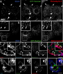
          The small GTPase Rab5 is a key regulator of clathrin-mediated endocytosis. On early endosomes, within a spatially restricted domain enriched in phosphatydilinositol-3-phosphate [PI(3)P], Rab5 coordinates a complex network of effectors that functionally cooperate in membrane tethering, fusion, and organelle motility. Here we discovered a novel PI(3)P-binding Rab5 effector, Rabankyrin-5, which localises to early endosomes and stimulates their fusion activity . In addition to early endosomes, however, Rabankyrin-5 localises to large vacuolar structures that correspond to macropinosomes in epithelial cells and fibroblasts. Overexpression of Rabankyrin-5 increases the number of macropinosomes and stimulates fluid-phase uptake, whereas its downregulation inhibits these processes. In polarised epithelial cells, this function is primarily restricted to the apical membrane. Rabankyrin-5 localises to large pinocytic structures underneath the apical surface of kidney proximal tubule cells, and its overexpression in polarised Madin-Darby canine kidney cells stimulates apical but not basolateral, non-clathrin-mediated pinocytosis. In demonstrating a regulatory role in endosome fusion and (macro)pinocytosis, our studies suggest that Rab5 regulates and coordinates different endocytic mechanisms through its effector Rabankyrin-5. Furthermore, its active role in apical pinocytosis in epithelial cells suggests an important function of Rabankyrin-5 in the physiology of polarised cells.

          Abstract

          Rabankyrin-5, which is activated by the small GTPase Rab5, coordinates two disparate methods that cells use to take in solids and fluids, playing a key role in both endosome fusion and macropinocytosis

          Related collections

          Most cited references89

          • Record: found
          • Abstract: found
          • Article: not found

          Regulated portals of entry into the cell.

          The plasma membrane is the interface between cells and their harsh environment. Uptake of nutrients and all communication among cells and between cells and their environment occurs through this interface. 'Endocytosis' encompasses several diverse mechanisms by which cells internalize macromolecules and particles into transport vesicles derived from the plasma membrane. It controls entry into the cell and has a crucial role in development, the immune response, neurotransmission, intercellular communication, signal transduction, and cellular and organismal homeostasis. As the complexity of molecular interactions governing endocytosis are revealed, it has become increasingly clear that it is tightly coordinated and coupled with overall cell physiology and thus, must be viewed in a broader context than simple vesicular trafficking.
            Bookmark
            • Record: found
            • Abstract: found
            • Article: not found

            A role for phosphoinositide 3-kinase in the completion of macropinocytosis and phagocytosis by macrophages

            Phosphoinositide 3-kinase (PI 3-kinase) has been implicated in growth factor signal transduction and vesicular membrane traffic. It is thought to mediate the earliest steps leading from ligation of cell surface receptors to increased cell surface ruffling. We show here that inhibitors of PI 3-kinase inhibit endocytosis in macrophages, not by interfering with the initiation of the process but rather by preventing its completion. Consistent with earlier studies, the inhibitors wortmannin and LY294002 inhibited fluid-phase pinocytosis and Fc receptor-mediated phagocytosis, but they had little effect on the receptor-mediated endocytosis of diI-labeled, acetylated, low density lipoprotein. Large solute probes of endocytosis reported greater inhibition by wortmannin than smaller probes did, indicating that macropinocytosis was affected more than micropinocytosis. Since macropinocytosis and phagocytosis are actin-mediated processes, we expected that their inhibition by wortmannin resulted from deficient signaling from macrophage colony-stimulating factor (M-CSF) receptors or Fc receptors to the actin cytoskeleton. However, video microscopy showed cell surface ruffling in wortmannin-treated cells, and increased ruffling after addition of M-CSF or phorbol myristate acetate. Quantitative measurements of video data reported slightly diminished ruffling in wortmannin-treated cells. Remarkably, the ruffles that formed in wortmannin-treated macrophages all receded into the cytoplasm without closing into macropinosomes. Similarly, wortmannin and LY294002 did not inhibit the extension of actin-rich pseudopodia along IgG- opsonized sheep erythrocytes, but instead prevented them from closing into phagosomes. These findings indicate that PI 3-kinase is not necessary for receptor-mediated stimulation of pseudopod extension, but rather functions in the closure of macropinosomes and phagosomes into intracellular organelles.
              Bookmark
              • Record: found
              • Abstract: found
              • Article: not found

              Distinct Membrane Domains on Endosomes in the Recycling Pathway Visualized by Multicolor Imaging of Rab4, Rab5, and Rab11

              Two endosome populations involved in recycling of membranes and receptors to the plasma membrane have been described, the early and the recycling endosome. However, this distinction is mainly based on the flow of cargo molecules and the spatial distribution of these membranes within the cell. To get insights into the membrane organization of the recycling pathway, we have studied Rab4, Rab5, and Rab11, three regulatory components of the transport machinery. Following transferrin as cargo molecule and GFP-tagged Rab proteins we could show that cargo moves through distinct domains on endosomes. These domains are occupied by different Rab proteins, revealing compartmentalization within the same continuous membrane. Endosomes are comprised of multiple combinations of Rab4, Rab5, and Rab11 domains that are dynamic but do not significantly intermix over time. Three major populations were observed: one that contains only Rab5, a second with Rab4 and Rab5, and a third containing Rab4 and Rab11. These membrane domains display differential pharmacological sensitivity, reflecting their biochemical and functional diversity. We propose that endosomes are organized as a mosaic of different Rab domains created through the recruitment of specific effector proteins, which cooperatively act to generate a restricted environment on the membrane.
                Bookmark

                Author and article information

                Journal
                PLoS Biol
                pbio
                PLoS Biology
                Public Library of Science (San Francisco, USA )
                1544-9173
                1545-7885
                September 2004
                24 August 2004
                : 2
                : 9
                : e261
                Affiliations
                [1] 1Max-Planck Institute of Molecular Cell Biology and Genetics DresdenGermany
                [2] 2Laboratory of Biological Chemistry, Medical School University of Ioannina, IoanninaGreece
                [3] 3Institute for Molecular Bioscience, Centre for Microscopy and Microanalysis School of Biomedical Sciences, University of Queensland, Brisbane, QueenslandAustralia
                [4] 4European Molecular Biology Laboratory HeidelbergGermany
                Article
                10.1371/journal.pbio.0020261
                514490
                15328530
                aa5356eb-c3f8-4411-a2c3-03adea1c77b1
                Copyright: © Schnatwinkel 2004 et al. This is an open-access article distributed under the terms of the Creative Commons Attribution License, which permits unrestricted use, distribution, and reproduction in any medium, provided the original work is properly cited
                History
                : 19 December 2003
                : 11 June 2004
                Categories
                Research Article
                Cell Biology
                Mammals

                Life sciences
                Life sciences

                Comments

                Comment on this article