11
views
0
recommends
+1 Recommend
0 collections
    0
    shares
      • Record: found
      • Abstract: found
      • Article: found
      Is Open Access

      Machine learning models based on quantitative dynamic contrast-enhanced MRI parameters assess the expression levels of CD3 +, CD4 +, and CD8 + tumor-infiltrating lymphocytes in advanced gastric carcinoma

      research-article

      Read this article at

      Bookmark
          There is no author summary for this article yet. Authors can add summaries to their articles on ScienceOpen to make them more accessible to a non-specialist audience.

          Abstract

          Objective

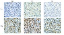
          To explore the effectiveness of machine learning classifiers based on dynamic contrast-enhanced magnetic resonance imaging (DCE-MRI) in predicting the expression levels of CD3 +, CD4 +, and CD8 + tumor-infiltrating lymphocytes (TILs) in patients with advanced gastric cancer (AGC).

          Methods

          This study investigated 103 patients with confirmed AGC through DCE-MRI and immunohistochemical staining. Immunohistochemical staining was used to evaluate CD3 +, CD4 +, and CD8 + T-cell expression. Utilizing Omni Kinetics software, radiomics features (K trans, K ep, and V e) were extracted and underwent selection via variance threshold, SelectKBest, and LASSO methods. Logistic regression (LR), support vector machine (SVM), random forest (RF), and eXtreme Gradient Boosting (XGBoost) are the four classifiers used to build four machine learning (ML) models, and their performance was evaluated using 10-fold cross-validation. The model’s performance was evaluated and compared using the area under the receiver operating characteristic curve (AUC), accuracy, sensitivity, specificity, positive predictive value, and negative predictive value.

          Results

          In terms of CD3 +, CD4 +, and CD8 + T lymphocyte prediction models, the random forest model outperformed the other classifier models in terms of CD4 + and CD8 + T cell prediction, with AUCs of 0.913 and 0.970 on the training set and 0.904 and 0.908 on the validation set, respectively. In terms of CD3 + T cell prediction, the logistic regression model fared the best, with AUCs on the training and validation sets of 0.872 and 0.817, respectively.

          Conclusion

          Machine learning classifiers based on DCE-MRI have the potential to accurately predict CD3 +, CD4 +, and CD8 + tumor-infiltrating lymphocyte expression levels in patients with AGC.

          Related collections

          Most cited references33

          • Record: found
          • Abstract: found
          • Article: not found

          Global cancer statistics 2020: GLOBOCAN estimates of incidence and mortality worldwide for 36 cancers in 185 countries

          This article provides an update on the global cancer burden using the GLOBOCAN 2020 estimates of cancer incidence and mortality produced by the International Agency for Research on Cancer. Worldwide, an estimated 19.3 million new cancer cases (18.1 million excluding nonmelanoma skin cancer) and almost 10.0 million cancer deaths (9.9 million excluding nonmelanoma skin cancer) occurred in 2020. Female breast cancer has surpassed lung cancer as the most commonly diagnosed cancer, with an estimated 2.3 million new cases (11.7%), followed by lung (11.4%), colorectal (10.0 %), prostate (7.3%), and stomach (5.6%) cancers. Lung cancer remained the leading cause of cancer death, with an estimated 1.8 million deaths (18%), followed by colorectal (9.4%), liver (8.3%), stomach (7.7%), and female breast (6.9%) cancers. Overall incidence was from 2-fold to 3-fold higher in transitioned versus transitioning countries for both sexes, whereas mortality varied <2-fold for men and little for women. Death rates for female breast and cervical cancers, however, were considerably higher in transitioning versus transitioned countries (15.0 vs 12.8 per 100,000 and 12.4 vs 5.2 per 100,000, respectively). The global cancer burden is expected to be 28.4 million cases in 2040, a 47% rise from 2020, with a larger increase in transitioning (64% to 95%) versus transitioned (32% to 56%) countries due to demographic changes, although this may be further exacerbated by increasing risk factors associated with globalization and a growing economy. Efforts to build a sustainable infrastructure for the dissemination of cancer prevention measures and provision of cancer care in transitioning countries is critical for global cancer control.
            Bookmark
            • Record: found
            • Abstract: found
            • Article: not found

            Understanding the tumor immune microenvironment (TIME) for effective therapy

            The clinical successes in immunotherapy have been both astounding and at the same time unsatisfactory. Countless patients with varied tumor types have seen pronounced clinical response with immunotherapeutic intervention; however, many more patients have experienced minimal or no clinical benefit when provided the same treatment. As technology has advanced, so has the understanding of the complexity and diversity of the immune context of the tumor microenvironment and its influence on response to therapy. It has been possible to identify different subclasses of immune environment that have an influence on tumor initiation and response and therapy; by parsing the unique classes and subclasses of tumor immune microenvironment (TIME) that exist within a patient’s tumor, the ability to predict and guide immunotherapeutic responsiveness will improve, and new therapeutic targets will be revealed.
              Bookmark
              • Record: found
              • Abstract: found
              • Article: not found

              Gastric cancer

              Gastric cancer is the fifth most common cancer and the third most common cause of cancer death globally. Risk factors for the condition include Helicobacter pylori infection, age, high salt intake, and diets low in fruit and vegetables. Gastric cancer is diagnosed histologically after endoscopic biopsy and staged using CT, endoscopic ultrasound, PET, and laparoscopy. It is a molecularly and phenotypically highly heterogeneous disease. The main treatment for early gastric cancer is endoscopic resection. Non-early operable gastric cancer is treated with surgery, which should include D2 lymphadenectomy (including lymph node stations in the perigastric mesentery and along the celiac arterial branches). Perioperative or adjuvant chemotherapy improves survival in patients with stage 1B or higher cancers. Advanced gastric cancer is treated with sequential lines of chemotherapy, starting with a platinum and fluoropyrimidine doublet in the first line; median survival is less than 1 year. Targeted therapies licensed to treat gastric cancer include trastuzumab (HER2-positive patients first line), ramucirumab (anti-angiogenic second line), and nivolumab or pembrolizumab (anti-PD-1 third line).
                Bookmark

                Author and article information

                Contributors
                URI : https://loop.frontiersin.org/people/2013937Role: Role: Role: Role: Role: Role:
                URI : https://loop.frontiersin.org/people/1407847Role: Role: Role: Role: Role: Role:
                Role: Role: Role: Role: Role:
                Role: Role: Role:
                Role: Role: Role:
                Role: Role: Role: Role: Role:
                Journal
                Front Oncol
                Front Oncol
                Front. Oncol.
                Frontiers in Oncology
                Frontiers Media S.A.
                2234-943X
                14 March 2024
                2024
                : 14
                : 1365550
                Affiliations
                [1] 1 Department of Radiology, Shaoxing People’s Hospital, Shaoxing Hospital, Zhejiang University School of Medicine , Shaoxing, China
                [2] 2 Department of Pathology, Shaoxing People’s Hospital, Shaoxing Hospital, Zhejiang University School of Medicine , Shaoxing, China
                Author notes

                Edited by: Francesco Sabbatino, University of Salerno, Italy

                Reviewed by: Marco Cascella, G. Pascale National Cancer Institute Foundation (IRCCS), Italy

                Zitong Lin, Nanjing University, China

                *Correspondence: Zengxin Lu, luzx777@ 123456163.com
                Article
                10.3389/fonc.2024.1365550
                10973004
                38549936
                a9af800c-4187-483a-8d91-95203c24e686
                Copyright © 2024 Huang, Li, Wang, Yang, Jin and Lu

                This is an open-access article distributed under the terms of the Creative Commons Attribution License (CC BY). The use, distribution or reproduction in other forums is permitted, provided the original author(s) and the copyright owner(s) are credited and that the original publication in this journal is cited, in accordance with accepted academic practice. No use, distribution or reproduction is permitted which does not comply with these terms.

                History
                : 04 January 2024
                : 29 February 2024
                Page count
                Figures: 6, Tables: 4, Equations: 3, References: 33, Pages: 12, Words: 5880
                Funding
                The author(s) declare that financial support was received for the research, authorship, and/or publication of this article. This work was supported by the General Project of Zhejiang Province Health Science and Technology Plan (Grant number, 2021KY1150, 2022KY1296, and 2023SKY035).
                Categories
                Oncology
                Original Research
                Custom metadata
                Cancer Immunity and Immunotherapy

                Oncology & Radiotherapy
                dynamic contrast-enhanced magnetic resonance imaging,advanced gastric carcinoma,machine learning,cd3+ ,cd4+ ,cd8+

                Comments

                Comment on this article