8
views
0
recommends
+1 Recommend
0 collections
    0
    shares
      • Record: found
      • Abstract: found
      • Article: found
      Is Open Access

      Anatomic Variations of the Right Portal Vein: Prevalence, Imaging Features, and Implications for Successful Transjugular Intrahepatic Portosystemic Shunt Creation

      other

      Read this article at

      Bookmark
          There is no author summary for this article yet. Authors can add summaries to their articles on ScienceOpen to make them more accessible to a non-specialist audience.

          Abstract

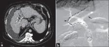
          Given the widespread use of transjugular intrahepatic portosystemic shunt (TIPS) creation for the treatment of portal hypertensive complications, a working knowledge of portal venous anatomy is critical for interventional radiologists. The right portal vein – which is most commonly accessed during TIPS – is subject to various anatomic variants that may potentially impact procedure success. This pictorial essay characterizes the anatomic patterns of the right portal vein branching in terms of type and frequency based on case series review. The work also explains the potential procedural implications of the right portal vein anatomic variations as they pertain to TIPS technical success.

          Related collections

          Most cited references12

          • Record: found
          • Abstract: found
          • Article: not found

          Insight into congenital absence of the portal vein: is it rare?

          Congenital absence of portal vein (CAPV) was a rare event in the past. However, the number of detected CAPV cases has increased in recent years because of advances in imaging techniques. Patients with CAPV present with portal hypertension (PH) or portosystemic encephalopathy (PSE), but these conditions rarely occur until the patients grow up or become old. The patients usually visit doctors for the complications of venous shunts, hepatic or cardiac abnormalities detected by ultrasonography (US), computed tomography (CT) and magnetic resonance imaging (MRI). The etiology of this disease is not clear, but most investigators consider that it is associated with abnormal embryologic development of the portal vein. Usually, surgical intervention can relieve the symptoms and prevent occurrence of complications in CAPV patients. Moreover, its management should be stressed on a case-by-case basis, depending on the type or anatomy of the disease, as well as the symptoms and clinical conditions of the patient.
            Bookmark
            • Record: found
            • Abstract: found
            • Article: not found

            Incidence, patterns, and clinical relevance of variant portal vein anatomy.

            The purpose of this study was to determine the incidence of variant intrahepatic portal vein anatomy detected on CT portography and to discuss surgical implications. Variant portal vein anatomy is nearly as common as variant hepatic artery anatomy. The complexity of hepatic interventions now performed by interventional radiologists and surgeons, including portal vein embolization, anatomic resection, and transplantation, make recognition and understanding of normal and variant portal vein anatomy increasingly important.
              Bookmark
              • Record: found
              • Abstract: not found
              • Article: not found

              Developmental basis of the portal venous system.

                Bookmark

                Author and article information

                Journal
                J Clin Imaging Sci
                J Clin Imaging Sci
                JCIS
                Journal of Clinical Imaging Science
                Medknow Publications & Media Pvt Ltd (India )
                2156-7514
                2156-5597
                2017
                28 March 2017
                : 7
                : 14
                Affiliations
                [1]Department of Radiology, Division of Interventional Radiology, University of Illinois Hospital and Health Sciences System, Chicago, IL 60612, USA
                Author notes
                Address for correspondence: Dr. Ron C Gaba, Department of Radiology, Division of Interventional Radiology, University of Illinois Hospital and Health Sciences System, 1740 West Taylor Street, MC 931, Chicago, IL 60612, USA. E-mail: rgaba@ 123456uic.edu
                Article
                JCIS-7-14
                10.4103/jcis.JCIS_10_17
                5385700
                28515965
                a9610e7c-6aad-41d2-acf6-864e48a5dea0
                Copyright: © 2017 Journal of Clinical Imaging Science

                This is an open access article distributed under the terms of the Creative Commons Attribution-NonCommercial-ShareAlike 3.0 License, which allows others to remix, tweak, and build upon the work non-commercially, as long as the author is credited and the new creations are licensed under the identical terms.

                History
                : 03 February 2017
                : 08 February 2017
                Categories
                Pictorial Essay

                Radiology & Imaging
                anatomy,portal vein,transjugular intrahepatic portosystemic shunt,variation

                Comments

                Comment on this article