1
views
0
recommends
+1 Recommend
0 collections
    0
    shares
      • Record: found
      • Abstract: found
      • Article: found
      Is Open Access

      Normal diameter of the optic nerve using magnetic resonance imaging: A retrospective Nigerian study

      other

      Read this article at

      Bookmark
          There is no author summary for this article yet. Authors can add summaries to their articles on ScienceOpen to make them more accessible to a non-specialist audience.

          Abstract

          PURPOSE:

          The variations in the diameter of the optic nerve (ON) are important clinically in the diagnosis of conditions associated with the ON such as raised intracranial pressure, meningioma, optic neuritis, and Grave’s orbitopathy. This study determined the normal diameters of the ON in adult Nigerians seen in a Hospital in Delta State.

          METHODS:

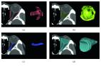
          Axial T1-weighted brain magnetic resonance imaging images of 150 patients (75 males and 75 females) aged ≥20 years were retrieved from the hospital’s radiological database and retrospectively used to evaluate the diameter of the ON on axial and coronal sections. The data were analyzed and summarized using descriptive statistics. The mean diameters were compared based on gender, side, and age groups and correlated with age using inferential statistics. The significance level was considered at 5%.

          RESULTS:

          The diameter of the ON measured 0.45 ± 0.07 cm on the coronal section, besides 0.50 ± 0.07 cm, and 0.46 ± 0.06 cm at 0.3 cm and 0.8 cm from the posterior pole of the globe, respectively, on the axial slices. The diameters were significantly larger in males than in females ( P < 0.05) and were symmetrical. However, they lacked significant association with age ( P > 0.05). The three diameters measured had a significant positive correlation with each other ( P < 0.05).

          CONCLUSION:

          The study provides a normal range of ON diameter in the study center to aid in the diagnosis of raised intracranial pressure and pathologies involving the nerve and its sheath.

          Related collections

          Most cited references16

          • Record: found
          • Abstract: found
          • Article: not found

          Normative measurements of orbital structures using CT.

          The purposes of this study were to establish criteria for the diameters of normal extraocular muscles, to determine the normal position of the globe as revealed by CT, and to investigate the effects of age and sex on these structures. Diameters of extraocular muscles, distance from the interzygomatic line to the posterior margin of the globe, width of the optic nerve-sheath complex, and length of the interzygomatic line were calculated for 200 normal orbits of 100 patients on axial and direct coronal CT images. Effects of age and sex on muscle diameters and globe position were analyzed. Normal ranges for the diameters (mean +/- 2SDs) of extraocular muscles were medial rectus, 3.3-5.0 mm; lateral rectus, 1.7-4.8 mm; inferior rectus, 3.2-6.5 mm; and superior group, 3.2-6.1 mm. The normal position of the globe was 9.4 mm behind the interzygomatic line (range, 5.9-12.8 mm). The mean diameters of the extraocular muscles and the length of the interzygomatic line in male patients were significantly larger than in female patients (p < .001). Statistically significant correlation was found between age and the diameters of the inferior and lateral rectus muscles (r = .32, p = .013; and r = .23, p = .048, respectively). Our results may be important in interpreting CT scans of the orbit because, to our knowledge, no reliable normative data exist regarding these orbital structures.
            Bookmark
            • Record: found
            • Abstract: found
            • Article: found
            Is Open Access

            Computed tomography imaging-based normative orbital measurement in Indian population

            Purpose: This study aims to evaluate normal orbital structures with nonenhanced computed tomography (NCCT) and determine normative data for the Indian population. Methods: CT images of the orbits of 100 patients were retrospectively reviewed on a work station to record the normative data of the orbits. Clinical details of all patients were reviewed to ensure that they did not have ocular/orbital diseases. Both axial and coronal images were utilized to record the data. Results: The mean age of the population evaluated was 34.07 years, with male to female ratio of 1.77. The average orbital index for the left orbit was 97 and for the right side was 103. The mean thickness of left inferior rectus, lateral rectus, medial rectus, and the superior rectus was 3.36 mm, 3.14 mm, 3.80 mm, and 3.75 mm, respectively. The right inferior rectus, lateral rectus, medial rectus, and the superior rectus measured 3.46 mm, 3.14 mm, 3.83 mm, and 3.78 mm, respectively. The optic nerve sheath complex diameter varied between 3.05 mm and 7.17 mm for the left eye and 3.05 mm and 7.0 mm for the right eye. Conclusion: The study provides normative data on various orbital structures in an Indian population. This data is likely to be useful for diagnosing various orbital pathologies and in planning surgical orbital procedures.
              Bookmark
              • Record: found
              • Abstract: found
              • Article: found
              Is Open Access

              Measurement of the Orbital Soft Tissue Volume in Chinese Adults Based on Three-Dimensional CT Reconstruction

              Quantitative measurement of the orbital soft tissue volume plays a very important role in the study of orbital diseases. The purpose of this study is to establish a computed tomography- (CT-) based three-dimensional (3D) reconstruction model and measure the orbital soft tissue volume in Chinese adults. We collected data from 103 Chinese adults (52 males and 51 females) who underwent orbital CT. The CT images of these adults were used to reconstruct a 3D model of the orbital bony cavity, orbital fat, extraocular muscle, and intraorbital optic nerve using Mimics software, and their respective volumes were measured. The mean (±SD) orbital bony cavity volume (OV), orbital fat volume (FV), extraocular muscle volume (MV), and intraorbital optic nerve volume (iONV) of the males were 22.2 ± 2.2 cm3, 8.9 ± 1.8 cm3, 1.9 ± 0.34 cm3, and 0.41 ± 0.08 cm3, respectively. The mean OV, FV, MV, and iONV of the females were 20.2 ± 1.5 cm3, 8.1 ± 1.7 cm3, 1.6 ± 0.3 cm3, and 0.36 ± 0.074 cm3, respectively, which were all significantly lower than those in males (all p < 0.05). FV (r = 0.370; p < 0.001) and MV (r = 0.283; p=0.007) were significantly correlated with body mass index (BMI), while iONV was not correlated with BMI (r = −0.070; p=0.480). This study shows that FV, MV, and iONV were higher in males than in females. With increasing BMI, FV and MV both increased, but iONV did not exhibit this trend.
                Bookmark

                Author and article information

                Journal
                Saudi J Ophthalmol
                Saudi J Ophthalmol
                SJO
                Saudi J Ophthalmol
                Saudi Journal of Ophthalmology
                Wolters Kluwer - Medknow (India )
                1319-4534
                2542-6680
                Jan-Mar 2024
                22 February 2024
                : 38
                : 1
                : 53-58
                Affiliations
                [1 ]Department of Human Anatomy and Cell Biology, Delta State University, Abraka, Nigeria
                [2 ]Department of Ophthalmology, Delta State University Teaching Hospital, Oghara, Nigeria
                [3 ]Department of Radiology, Delta State University Teaching Hospital, Oghara, Nigeria
                Author notes
                Address for correspondence: Dr. Joyce E. Ikubor, Department of Radiology, Delta State University Teaching Hospital, Oghara, Nigeria. E-mail: joyceikuborjune12@ 123456yahoo.com
                Article
                SJO-38-53
                10.4103/sjopt.sjopt_189_23
                11017001
                38628409
                a8882c7b-2308-423f-ae2e-4edd70f5c290
                Copyright: © 2024 Saudi Journal of Ophthalmology

                This is an open access journal, and articles are distributed under the terms of the Creative Commons Attribution-NonCommercial-ShareAlike 4.0 License, which allows others to remix, tweak, and build upon the work non-commercially, as long as appropriate credit is given and the new creations are licensed under the identical terms.

                History
                : 08 August 2023
                : 21 November 2023
                : 25 December 2023
                Categories
                Neuro-ophthalmology Update

                diameter,magnetic resonance imaging,optic nerve,sheath
                diameter, magnetic resonance imaging, optic nerve, sheath

                Comments

                Comment on this article