2
views
0
recommends
+1 Recommend
0 collections
    0
    shares
      • Record: found
      • Abstract: found
      • Article: found
      Is Open Access

      Towards the Segmentation and Classification of White Blood Cell Cancer Using Hybrid Mask-Recurrent Neural Network and Transfer Learning

      research-article

      Read this article at

      Bookmark
          There is no author summary for this article yet. Authors can add summaries to their articles on ScienceOpen to make them more accessible to a non-specialist audience.

          Abstract

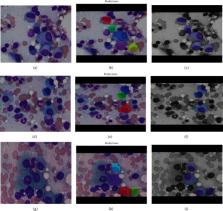
          Inside the bone marrow, plasma cells are created, and they are a type of white blood cells. They are made from B lymphocytes. Antigens are produced by plasma cells to combat bacteria and viruses and prevent inflammation and illness. Multiple myeloma is a plasma cell cancer that starts in the bone marrow and causes the formation of abnormal plasma cells. Multiple myeloma is firmly identified by examining bone marrow samples under a microscope for myeloma cells. To diagnose myeloma cells, pathologists have to be very selective. Furthermore, because the ultimate decision is based on human sight and opinion, there is a possibility of error in the result. The nobility of this research is that it provides a computer-assisted technique for recognizing and detecting myeloma cells in bone marrow smears. For recognizing purposes, we have used Mask-Recurrent Convolutional Neural Network, and for detection purposes, Efficient Net B3 has been used. There are already many studies on white blood cell cancer, but very few with both segmentation and classification. We have designed two models. One is for recognizing myeloma cells, and the other is for differentiating them from nonmyeloma cells. Also, a new data set has been made from the multiple myeloma data sets, which has been used in our classification model. This research focuses on hybrid segmentation models and increases the accuracy level of the classification model. Both of our models are trained pretty well, where the Mask-RCNN model gives a mean average precision (mAP) of 93% and the Efficient Net B3 model gives 94.68% accuracy. The result of this research indicates that the Mask-RCNN model can recognize multiple myeloma and Efficient Net B3 can distinguish between myeloma and nonmyeloma cells and beats most of the state of the art in myeloma recognition and detection.

          Related collections

          Most cited references26

          • Record: found
          • Abstract: not found
          • Conference Proceedings: not found

          Deep Residual Learning for Image Recognition

            Bookmark
            • Record: found
            • Abstract: found
            • Article: found

            Deep Learning in Medical Image Analysis

            This review covers computer-assisted analysis of images in the field of medical imaging. Recent advances in machine learning, especially with regard to deep learning, are helping to identify, classify, and quantify patterns in medical images. At the core of these advances is the ability to exploit hierarchical feature representations learned solely from data, instead of features designed by hand according to domain-specific knowledge. Deep learning is rapidly becoming the state of the art, leading to enhanced performance in various medical applications. We introduce the fundamentals of deep learning methods and review their successes in image registration, detection of anatomical and cellular structures, tissue segmentation, computer-aided disease diagnosis and prognosis, and so on. We conclude by discussing research issues and suggesting future directions for further improvement.
              Bookmark
              • Record: found
              • Abstract: found
              • Article: found
              Is Open Access

              Automated Whole-Body Bone Lesion Detection for Multiple Myeloma on 68 Ga-Pentixafor PET/CT Imaging Using Deep Learning Methods

              The identification of bone lesions is crucial in the diagnostic assessment of multiple myeloma (MM). 68Ga-Pentixafor PET/CT can capture the abnormal molecular expression of CXCR-4 in addition to anatomical changes. However, whole-body detection of dozens of lesions on hybrid imaging is tedious and error prone. It is even more difficult to identify lesions with a large heterogeneity. This study employed deep learning methods to automatically combine characteristics of PET and CT for whole-body MM bone lesion detection in a 3D manner. Two convolutional neural networks (CNNs), V-Net and W-Net, were adopted to segment and detect the lesions. The feasibility of deep learning for lesion detection on 68Ga-Pentixafor PET/CT was first verified on digital phantoms generated using realistic PET simulation methods. Then the proposed methods were evaluated on real 68Ga-Pentixafor PET/CT scans of MM patients. The preliminary results showed that deep learning method can leverage multimodal information for spatial feature representation, and W-Net obtained the best result for segmentation and lesion detection. It also outperformed traditional machine learning methods such as random forest classifier (RF), k-Nearest Neighbors (k-NN), and support vector machine (SVM). The proof-of-concept study encourages further development of deep learning approach for MM lesion detection in population study.
                Bookmark

                Author and article information

                Contributors
                Journal
                Contrast Media Mol Imaging
                Contrast Media Mol Imaging
                CMMI
                Contrast Media & Molecular Imaging
                Hindawi
                1555-4309
                1555-4317
                2021
                2 December 2021
                : 2021
                : 4954854
                Affiliations
                1Department of Electrical and Computer Engineering, North South University, Dhaka-1229, Bangladesh
                2Department of Information Technology, College of Computers and Information Technology, Taif University, P.O. Box 11099, Taif 21944, Saudi Arabia
                Author notes

                Academic Editor: Yuvaraja Teekaraman

                Author information
                https://orcid.org/0000-0001-8008-6921
                https://orcid.org/0000-0002-3844-8631
                https://orcid.org/0000-0002-0200-975X
                https://orcid.org/0000-0003-0779-8820
                https://orcid.org/0000-0002-6638-7039
                Article
                10.1155/2021/4954854
                8660215
                a73caefb-9659-4836-a4e1-a7e748f92266
                Copyright © 2021 Sumit Kumar Das et al.

                This is an open access article distributed under the Creative Commons Attribution License, which permits unrestricted use, distribution, and reproduction in any medium, provided the original work is properly cited.

                History
                : 11 October 2021
                : 19 November 2021
                Funding
                Funded by: Taif University
                Award ID: TURSP-2020/26
                Categories
                Research Article

                Comments

                Comment on this article