24
views
0
recommends
+1 Recommend
0 collections
    0
    shares
      • Record: found
      • Abstract: found
      • Article: found
      Is Open Access

      Collagen Promotes Higher Adhesion, Survival and Proliferation of Mesenchymal Stem Cells

      research-article

      Read this article at

      Bookmark
          There is no author summary for this article yet. Authors can add summaries to their articles on ScienceOpen to make them more accessible to a non-specialist audience.

          Abstract

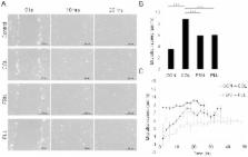
          Mesenchymal stem cells (MSC) can differentiate into several cell types and are desirable candidates for cell therapy and tissue engineering. However, due to poor cell survival, proliferation and differentiation in the patient, the therapy outcomes have not been satisfactory. Although several studies have been done to understand the conditions that promote proliferation, differentiation and migration of MSC in vitro and in vivo, still there is no clear understanding on the effect of non-cellular bio molecules. Of the many factors that influence the cell behavior, the immediate cell microenvironment plays a major role. In this context, we studied the effect of extracellular matrix (ECM) proteins in controlling cell survival, proliferation, migration and directed MSC differentiation. We found that collagen promoted cell proliferation, cell survival under stress and promoted high cell adhesion to the cell culture surface. Increased osteogenic differentiation accompanied by high active RHOA (Ras homology gene family member A) levels was exhibited by MSC cultured on collagen. In conclusion, our study shows that collagen will be a suitable matrix for large scale production of MSC with high survival rate and to obtain high osteogenic differentiation for therapy.

          Related collections

          Most cited references34

          • Record: found
          • Abstract: found
          • Article: not found

          The control of human mesenchymal cell differentiation using nanoscale symmetry and disorder.

          A key tenet of bone tissue engineering is the development of scaffold materials that can stimulate stem cell differentiation in the absence of chemical treatment to become osteoblasts without compromising material properties. At present, conventional implant materials fail owing to encapsulation by soft tissue, rather than direct bone bonding. Here, we demonstrate the use of nanoscale disorder to stimulate human mesenchymal stem cells (MSCs) to produce bone mineral in vitro, in the absence of osteogenic supplements. This approach has similar efficiency to that of cells cultured with osteogenic media. In addition, the current studies show that topographically treated MSCs have a distinct differentiation profile compared with those treated with osteogenic media, which has implications for cell therapies.
            Bookmark
            • Record: found
            • Abstract: found
            • Article: not found

            Adult mesenchymal stem cells for tissue engineering versus regenerative medicine.

            Adult mesenchymal stem cells (MSCs) can be isolated from bone marrow or marrow aspirates and because they are culture-dish adherent, they can be expanded in culture while maintaining their multipotency. The MSCs have been used in preclinical models for tissue engineering of bone, cartilage, muscle, marrow stroma, tendon, fat, and other connective tissues. These tissue-engineered materials show considerable promise for use in rebuilding damaged or diseased mesenchymal tissues. Unanticipated is the realization that the MSCs secrete a large spectrum of bioactive molecules. These molecules are immunosuppressive, especially for T-cells and, thus, allogeneic MSCs can be considered for therapeutic use. In this context, the secreted bioactive molecules provide a regenerative microenvironment for a variety of injured adult tissues to limit the area of damage and to mount a self-regulated regenerative response. This regenerative microenvironment is referred to as trophic activity and, therefore, MSCs appear to be valuable mediators for tissue repair and regeneration. The natural titers of MSCs that are drawn to sites of tissue injury can be augmented by allogeneic MSCs delivered via the bloodstream. Indeed, human clinical trials are now under way to use allogeneic MSCs for treatment of myocardial infarcts, graft-versus-host disease, Crohn's Disease, cartilage and meniscus repair, stroke, and spinal cord injury. This review summarizes the biological basis for the in vivo functioning of MSCs through development and aging.
              Bookmark
              • Record: found
              • Abstract: found
              • Article: not found

              Geometric cues for directing the differentiation of mesenchymal stem cells.

              Significant efforts have been directed to understanding the factors that influence the lineage commitment of stem cells. This paper demonstrates that cell shape, independent of soluble factors, has a strong influence on the differentiation of human mesenchymal stem cells (MSCs) from bone marrow. When exposed to competing soluble differentiation signals, cells cultured in rectangles with increasing aspect ratio and in shapes with pentagonal symmetry but with different subcellular curvature-and with each occupying the same area-display different adipogenesis and osteogenesis profiles. The results reveal that geometric features that increase actomyosin contractility promote osteogenesis and are consistent with in vivo characteristics of the microenvironment of the differentiated cells. Cytoskeletal-disrupting pharmacological agents modulate shape-based trends in lineage commitment verifying the critical role of focal adhesion and myosin-generated contractility during differentiation. Microarray analysis and pathway inhibition studies suggest that contractile cells promote osteogenesis by enhancing c-Jun N-terminal kinase (JNK) and extracellular related kinase (ERK1/2) activation in conjunction with elevated wingless-type (Wnt) signaling. Taken together, this work points to the role that geometric shape cues can play in orchestrating the mechanochemical signals and paracrine/autocrine factors that can direct MSCs to appropriate fates.
                Bookmark

                Author and article information

                Contributors
                Role: Editor
                Journal
                PLoS One
                PLoS ONE
                plos
                plosone
                PLoS ONE
                Public Library of Science (San Francisco, CA USA )
                1932-6203
                14 December 2015
                2015
                : 10
                : 12
                : e0145068
                Affiliations
                [1 ]Stem Cell Biology Lab, Department of Biosciences and Bioengineering, Indian Institute of Technology Guwahati, Guwahati, Assam, India
                [2 ]Department of Biosciences and Bioengineering, Indian Institute of Technology Guwahati, Guwahati, Assam, India
                [3 ]Department of Hematology, Gauhati Medical College Hospital, Assam, India
                Rutgers - New Jersey Medical School, UNITED STATES
                Author notes

                Competing Interests: The authors have declared that no competing interests exist.

                Conceived and designed the experiments: BGJ AK CS. Performed the experiments: CS AK DM. Analyzed the data: BGJ AK CS AS SDP. Contributed reagents/materials/analysis tools: JB RS. Wrote the paper: BGJ AK JB CS.

                Article
                PONE-D-15-35153
                10.1371/journal.pone.0145068
                4678765
                26661657
                a71dedbb-c06f-4d47-97fa-f5e42627554f
                © 2015 Somaiah et al

                This is an open access article distributed under the terms of the Creative Commons Attribution License, which permits unrestricted use, distribution, and reproduction in any medium, provided the original author and source are credited

                History
                : 11 August 2015
                : 29 November 2015
                Page count
                Figures: 5, Tables: 0, Pages: 15
                Funding
                CS, AK, DM are supported by fellowship from Ministry of Human Resource Development, Govt. of India. This work was supported by funds from Department of Science and Technology SR/FT/LS-97/2009 (DST), Govt. of India and Department of Biotechnology (DBT), Govt. of India under RGYI (Rapid Grant for Young Investigator) scheme BT/PRI/5091/GBD/27/310/2011 to BGJ ( http://www.dbtindia.nic.in/). TIRF measurement was performed in facility supported by DBT to RS. The funders had no role in study design, data collection and analysis, decision to publish, or preparation of the manuscript.
                Categories
                Research Article
                Custom metadata
                All relevant data are within the paper and its Supporting Information files.

                Uncategorized
                Uncategorized

                Comments

                Comment on this article