4
views
0
recommends
+1 Recommend
0 collections
    0
    shares
      • Record: found
      • Abstract: found
      • Article: found
      Is Open Access

      17β-estradiol-induced mitochondrial dysfunction and Warburg effect in cervical cancer cells allow cell survival under metabolic stress

      research-article

      Read this article at

      Bookmark
          There is no author summary for this article yet. Authors can add summaries to their articles on ScienceOpen to make them more accessible to a non-specialist audience.

          Abstract

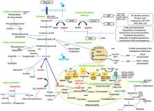
          Mitochondria from different types of cancer show bioenergetics and dysfunction that favor cell proliferation. The mechanistic understanding of estrogen in cervical cancer is poorly understood. Therefore, the objective of this study was to determine how 17β-estradiol (E2) affects mitochondrial function and the Warburg effect in SiHa, HeLa and C33A cervical cancer cells. Mitochondrial compromise was evaluated measuring changes in the membrane permeability by immunofluorescence, calcium concentration, redox status, iron and ferritin reserves. Glucose consumption and lactic acid assays were used to detect the metabolic activity. Results were confirmed at molecular level by analysis of the differential gene expression using RNA sequencing. E2 modified the mitochondrial permeability and produced an alteration in the calcium signaling pathway. In HeLa and SiHa, there was a significant decrease in nitric oxide levels and lipid peroxidation, and an increase in glucose consumption and lactic acid levels when stimulated with E2. Intracellular iron or ferritin reserves were not affected by the E2 treatment. Genes differentially modulated by E2 were involved in the mitochondrial electron transport chain, oxidative phosphorylation system, glycolysis, pentose phosphate pathway and the regulation of metabolic signaling pathways. Herein, we provide evidence for a primary effect of estrogen on mitochondrial function and the Warburg effect, favoring the metabolic adaptation of the cervical cancer cell lines and their survival.

          Related collections

          Most cited references57

          • Record: found
          • Abstract: found
          • Article: not found

          AMPK is a negative regulator of the Warburg effect and suppresses tumor growth in vivo.

          AMPK is a metabolic sensor that helps maintain cellular energy homeostasis. Despite evidence linking AMPK with tumor suppressor functions, the role of AMPK in tumorigenesis and tumor metabolism is unknown. Here we show that AMPK negatively regulates aerobic glycolysis (the Warburg effect) in cancer cells and suppresses tumor growth in vivo. Genetic ablation of the α1 catalytic subunit of AMPK accelerates Myc-induced lymphomagenesis. Inactivation of AMPKα in both transformed and nontransformed cells promotes a metabolic shift to aerobic glycolysis, increased allocation of glucose carbon into lipids, and biomass accumulation. These metabolic effects require normoxic stabilization of the hypoxia-inducible factor-1α (HIF-1α), as silencing HIF-1α reverses the shift to aerobic glycolysis and the biosynthetic and proliferative advantages conferred by reduced AMPKα signaling. Together our findings suggest that AMPK activity opposes tumor development and that its loss fosters tumor progression in part by regulating cellular metabolic pathways that support cell growth and proliferation. Copyright © 2013 Elsevier Inc. All rights reserved.
            Bookmark
            • Record: found
            • Abstract: found
            • Article: not found

            Ferroptosis: Role of lipid peroxidation, iron and ferritinophagy.

            Ferroptosis is a form of regulated cell death that is dependent on iron and reactive oxygen species (ROS) and is characterized by lipid peroxidation. It is morphologically and biochemically distinct and disparate from other processes of cell death. As ferroptosis is induced by inhibition of cysteine uptake or inactivation of the lipid repair enzyme glutathione peroxidase 4 (GPX4), the process is favored by chemical or mutational inhibition of the cystine/glutamate antiporter and culminates in the accumulation of reactive oxygen species (ROS) in the form of lipid hydroperoxides. Excessive lipid peroxidation leads to death by ferroptosis and the phenotype is accentuated respectively by the repletion and depletion of iron and glutathione in cells. Furthermore, oxidized phosphatidylethanolamines (PE) harbouring arachidonoyl (AA) and adrenoyl moieties (AdA) have been shown as proximate executioners of ferroptosis. Induction of ferroptosis due to cysteine depletion leads to the degradation of ferritin (i.e. ferritinophagy), which releases iron via the NCOA4-mediated autophagy pathway. Evidence of the manifestation of ferroptosis in vivo in iron overload mice mutants is emerging. Thus, a concerted synchronization of iron availability, ROS generation, glutamate excess and cysteine deficit leads to ferroptosis. A number of questions on the molecular mechanisms of some features of ferroptosis are highlighted as subjects for future investigations.
              Bookmark
              • Record: found
              • Abstract: found
              • Article: not found

              Estrogen receptors alpha (ERα) and beta (ERβ): subtype-selective ligands and clinical potential.

              Estrogen receptors alpha (ERα) and beta (ERβ) are nuclear transcription factors that are involved in the regulation of many complex physiological processes in humans. Modulation of these receptors by prospective therapeutic agents is currently being considered for prevention and treatment of a wide variety of pathological conditions, such as, cancer, metabolic and cardiovascular diseases, neurodegeneration, inflammation, and osteoporosis. This review provides an overview and update of compounds that have been recently reported as modulators of ERs, with a particular focus on their potential clinical applications. Copyright © 2014 Elsevier Ltd. All rights reserved.
                Bookmark

                Author and article information

                Journal
                Int J Oncol
                Int. J. Oncol
                IJO
                International Journal of Oncology
                D.A. Spandidos
                1019-6439
                1791-2423
                January 2020
                14 November 2019
                14 November 2019
                : 56
                : 1
                : 33-46
                Affiliations
                [1 ]Laboratory of Immunology, Department of Physiology, University Center for Health Sciences, University of Guadalajara
                [2 ]Division of Immunology, Western Biomedical Research Center, Mexican Social Security Institute
                [3 ]Research Institute in Biomedical Sciences
                [4 ]Diagnostic Laboratory
                [5 ]Department of Neurosciences, University Center for Health Sciences, University of Guadalajara, Guadalajara, Jalisco 44340, Mexico
                Author notes
                Correspondence to: Dr Ana Laura Pereira-Suárez, Laboratory of Immunology, Department of Physiology, University Center for Health Sciences, University of Guadalajara, 950 Sierra Mojada, Colonia Independencia, Guadalajara, Jalisco 44340, Mexico, E-mail: analauraps@ 123456hotmail.com
                [*]

                Contributed equally

                Article
                ijo-56-01-0033
                10.3892/ijo.2019.4912
                6910176
                31746421
                a70a8b6c-6802-450c-bc1d-5cc9ed048993
                Copyright: © Leal et al.

                This is an open access article distributed under the terms of the Creative Commons Attribution-NonCommercial-NoDerivs License, which permits use and distribution in any medium, provided the original work is properly cited, the use is non-commercial and no modifications or adaptations are made.

                History
                : 04 April 2019
                : 19 August 2019
                Categories
                Articles

                mitochondrial function,antioxidant,warburg effect,estradiol

                Comments

                Comment on this article