- Record: found
- Abstract: found
- Article: found
Cone-beam computed tomographic analysis of middle mesial canals and isthmus in mesial roots of mandibular first molars-prevalence and related factors
Read this article at
Abstract
Context:
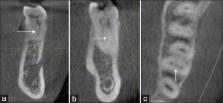
The internal anatomy of mandibular first molars has been studied in depth in different populations. However, limited information is present in differentiating a true middle mesial (MM) canal from an isthmus.
Aims:
The primary aim of this study was to identify the prevalence of a true MM canal and isthmus by retrospectively analyzing cone-beam computed tomography (CBCT) images in vivo. The secondary aim was to determine any correlation between related factors such as sex and age.
Materials and Methods:
CBCT images of 130 patients with the age group of 13–70 years were selected. Findings of MM canals and isthmus were recorded along with variables such as age and sex. Prevalence was compared using the Chi-square test ( P < 0.05).
Results:
Out of 143 mandibular first molars, the prevalence of the MM canal was 18.2%. There was no statistically significant difference between sex and prevalence of the MM canal and isthmus. The prevalence of isthmi in the mesial roots was 78.4%. Their presence was significantly higher in the apical third area (37.1%) ( P < 0.05). Both MM canal and isthmus were seen significantly higher in the age group of 31–50 years ( P < 0.05).
Related collections
Most cited references23
- Record: found
- Abstract: found
- Article: not found
Analysis of the internal anatomy of maxillary first molars by using different methods.
- Record: found
- Abstract: found
- Article: not found
Use of cone-beam computed tomography to evaluate root and canal morphology of mandibular molars in Chinese individuals.
- Record: found
- Abstract: found
- Article: not found