23
views
0
recommends
+1 Recommend
0 collections
    0
    shares
      • Record: found
      • Abstract: found
      • Article: found
      Is Open Access

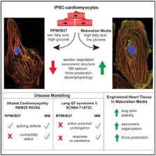
      Metabolic Maturation Media Improve Physiological Function of Human iPSC-Derived Cardiomyocytes

      research-article

      Read this article at

      Bookmark
          There is no author summary for this article yet. Authors can add summaries to their articles on ScienceOpen to make them more accessible to a non-specialist audience.

          SUMMARY

          Induced pluripotent stem cell-derived cardiomyocytes (iPSC-CMs) have enormous potential for the study of human cardiac disorders. However, their physiological immaturity severely limits their utility as a model system and their adoption for drug discovery. Here, we describe maturation media designed to provide oxidative substrates adapted to the metabolic needs of human iPSC (hiPSC)-CMs. Compared with conventionally cultured hiPSC-CMs, metabolically matured hiPSC-CMs contract with greater force and show an increased reliance on cardiac sodium (Na +) channels and sarcoplasmic reticulum calcium (Ca 2+) cycling. The media enhance the function, long-term survival, and sarcomere structures in engineered heart tissues. Use of the maturation media made it possible to reliably model two genetic cardiac diseases: long QT syndrome type 3 due to a mutation in the cardiac Na + channel SCN5A and dilated cardiomyopathy due to a mutation in the RNA splicing factor RBM20. The maturation media should increase the fidelity of hiPSC-CMs as disease models.

          Graphical Abstract

          In Brief

          Physiological immaturity of iPSC-derived cardiomyocytes limits their fidelity as disease models. Feyen et al. developed a low glucose, high oxidative substrate media that increase maturation of ventricular-like hiPSC-CMs in 2D and 3D cultures relative to standard protocols. Improved characteristics include a low resting V m, rapid depolarization, and increased Ca 2+ dependence and force generation.

          Related collections

          Most cited references38

          • Record: found
          • Abstract: found
          • Article: not found

          Cardiac metabolism and its interactions with contraction, growth, and survival of cardiomyocytes.

          The network for cardiac fuel metabolism contains intricate sets of interacting pathways that result in both ATP-producing and non-ATP-producing end points for each class of energy substrates. The most salient feature of the network is the metabolic flexibility demonstrated in response to various stimuli, including developmental changes and nutritional status. The heart is also capable of remodeling the metabolic pathways in chronic pathophysiological conditions, which results in modulations of myocardial energetics and contractile function. In a quest to understand the complexity of the cardiac metabolic network, pharmacological and genetic tools have been engaged to manipulate cardiac metabolism in a variety of research models. In concert, a host of therapeutic interventions have been tested clinically to target substrate preference, insulin sensitivity, and mitochondrial function. In addition, the contribution of cellular metabolism to growth, survival, and other signaling pathways through the production of metabolic intermediates has been increasingly noted. In this review, we provide an overview of the cardiac metabolic network and highlight alterations observed in cardiac pathologies as well as strategies used as metabolic therapies in heart failure. Lastly, the ability of metabolic derivatives to intersect growth and survival are also discussed.
            Bookmark
            • Record: found
            • Abstract: found
            • Article: not found

            Thyroid and Glucocorticoid Hormones Promote Functional T-Tubule Development in Human-Induced Pluripotent Stem Cell-Derived Cardiomyocytes.

            Human-induced pluripotent stem cell-derived cardiomyocytes (hiPSC-CM) are increasingly being used for modeling heart disease and are under development for regeneration of the injured heart. However, incomplete structural and functional maturation of hiPSC-CM, including lack of T-tubules, immature excitation-contraction coupling, and inefficient Ca-induced Ca release remain major limitations.
              Bookmark
              • Record: found
              • Abstract: found
              • Article: found
              Is Open Access

              Fatty Acids Enhance the Maturation of Cardiomyocytes Derived from Human Pluripotent Stem Cells

              Summary Although human pluripotent stem cell-derived cardiomyocytes (hPSC-CMs) have emerged as a novel platform for heart regeneration, disease modeling, and drug screening, their immaturity significantly hinders their application. A hallmark of postnatal cardiomyocyte maturation is the metabolic substrate switch from glucose to fatty acids. We hypothesized that fatty acid supplementation would enhance hPSC-CM maturation. Fatty acid treatment induces cardiomyocyte hypertrophy and significantly increases cardiomyocyte force production. The improvement in force generation is accompanied by enhanced calcium transient peak height and kinetics, and by increased action potential upstroke velocity and membrane capacitance. Fatty acids also enhance mitochondrial respiratory reserve capacity. RNA sequencing showed that fatty acid treatment upregulates genes involved in fatty acid β-oxidation and downregulates genes in lipid synthesis. Signal pathway analyses reveal that fatty acid treatment results in phosphorylation and activation of multiple intracellular kinases. Thus, fatty acids increase human cardiomyocyte hypertrophy, force generation, calcium dynamics, action potential upstroke velocity, and oxidative capacity. This enhanced maturation should facilitate hPSC-CM usage for cell therapy, disease modeling, and drug/toxicity screens.
                Bookmark

                Author and article information

                Journal
                101573691
                39703
                Cell Rep
                Cell Rep
                Cell reports
                2211-1247
                24 July 2020
                21 July 2020
                19 August 2020
                : 32
                : 3
                : 107925
                Affiliations
                [1 ]Cardiovascular Institute and Department of Medicine, Stanford University, Stanford, CA 94305, USA
                [2 ]Department of Cardiology, University Medical Center Utrecht, Utrecht, the Netherlands
                [3 ]Sanford-Burnham-Prebys Medical Discovery Institute, La Jolla, CA, USA
                [4 ]Department of Bioengineering, University of California, San Diego, San Diego, CA, USA
                [5 ]Institute of Experimental Pharmacology and Toxicology, University Medical Center Hamburg-Eppendorf, Hamburg, Germany
                [6 ]Electron Microscopy Unit, Center for Molecular Neurobiology Hamburg, University Medical Center Hamburg-Eppendorf, Hamburg, Germany
                [7 ]Department of Pharmacology, University of California, Davis, Davis, CA, USA
                [8 ]These authors contributed equally
                [9 ]Lead Contact
                Author notes

                AUTHOR CONTRIBUTIONS

                Conceptualization, D.A.M.F., W.L.M., A.A.N.B., H.Z., S.S., C.M.M., and M.M.; Methodology, D.A.M.F., W.L.M., A.A.N.B., and M.M.; Investigation, D.A.M.F., W.L.M., A.A.N.B., S.S., L.H., B.U., F.B., M.S., B.H., Z.L., R.-P.P., K.S.G., C.K.L., R.S., C.W., A.K., M.V., P.L.A., C.S.B., S.R., R.G.C.M., and M.G.; Writing–Original Draft, D.A.M.F., W.L.M., A.A.N.B., and M.M.; Writing–Review & Editing, all authors; Funding Acquisition, D.A.M.F., C.W., D.B., J.C.W., D.M.B., T.E., C.M.M., and M.M.; Supervision, J.C.W., D.M.B., T.E., and M.M.

                [* ]Correspondence: mmercola@ 123456stanford.edu
                Article
                NIHMS1613944
                10.1016/j.celrep.2020.107925
                7437654
                32697997
                a6e5209f-cccb-40fc-88d7-11f3bca80e82

                This is an open access article under the CC BY-NC-ND license ( http://creativecommons.org/licenses/by-nc-nd/4.0/).

                History
                Categories
                Article

                Cell biology
                Cell biology

                Comments

                Comment on this article