29
views
0
recommends
+1 Recommend
0 collections
    0
    shares
      • Record: found
      • Abstract: found
      • Article: found
      Is Open Access

      Solution Structure of an Archaeal DNA Binding Protein with an Eukaryotic Zinc Finger Fold

      research-article

      Read this article at

      Bookmark
          There is no author summary for this article yet. Authors can add summaries to their articles on ScienceOpen to make them more accessible to a non-specialist audience.

          Abstract

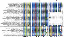
          While the basal transcription machinery in archaea is eukaryal-like, transcription factors in archaea and their viruses are usually related to bacterial transcription factors. Nevertheless, some of these organisms show predicted classical zinc fingers motifs of the C2H2 type, which are almost exclusively found in proteins of eukaryotes and most often associated with transcription regulators. In this work, we focused on the protein AFV1p06 from the hyperthermophilic archaeal virus AFV1. The sequence of the protein consists of the classical eukaryotic C2H2 motif with the fourth histidine coordinating zinc missing, as well as of N- and C-terminal extensions. We showed that the protein AFV1p06 binds zinc and solved its solution structure by NMR. AFV1p06 displays a zinc finger fold with a novel structure extension and disordered N- and C-termini. Structure calculations show that a glutamic acid residue that coordinates zinc replaces the fourth histidine of the C2H2 motif. Electromobility gel shift assays indicate that the protein binds to DNA with different affinities depending on the DNA sequence. AFV1p06 is the first experimentally characterised archaeal zinc finger protein with a DNA binding activity. The AFV1p06 protein family has homologues in diverse viruses of hyperthermophilic archaea. A phylogenetic analysis points out a common origin of archaeal and eukaryotic C2H2 zinc fingers.

          Related collections

          Most cited references44

          • Record: found
          • Abstract: found
          • Article: not found

          Protein backbone angle restraints from searching a database for chemical shift and sequence homology.

          Chemical shifts of backbone atoms in proteins are exquisitely sensitive to local conformation, and homologous proteins show quite similar patterns of secondary chemical shifts. The inverse of this relation is used to search a database for triplets of adjacent residues with secondary chemical shifts and sequence similarity which provide the best match to the query triplet of interest. The database contains 13C alpha, 13C beta, 13C', 1H alpha and 15N chemical shifts for 20 proteins for which a high resolution X-ray structure is available. The computer program TALOS was developed to search this database for strings of residues with chemical shift and residue type homology. The relative importance of the weighting factors attached to the secondary chemical shifts of the five types of resonances relative to that of sequence similarity was optimized empirically. TALOS yields the 10 triplets which have the closest similarity in secondary chemical shift and amino acid sequence to those of the query sequence. If the central residues in these 10 triplets exhibit similar phi and psi backbone angles, their averages can reliably be used as angular restraints for the protein whose structure is being studied. Tests carried out for proteins of known structure indicate that the root-mean-square difference (rmsd) between the output of TALOS and the X-ray derived backbone angles is about 15 degrees. Approximately 3% of the predictions made by TALOS are found to be in error.
            Bookmark
            • Record: found
            • Abstract: found
            • Article: not found

            Backbone dynamics of a free and phosphopeptide-complexed Src homology 2 domain studied by 15N NMR relaxation.

            The backbone dynamics of the C-terminal SH2 domain of phospholipase C gamma 1 have been investigated. Two forms of the domain were studied, one in complex with a high-affinity binding peptide derived from the platelet-derived growth factor receptor and the other in the absence of this peptide. 2-D 1H-15N NMR methods, employing pulsed field gradients, were used to determine steady-state 1H-15N NOE values and T1 and T2 15N relaxation times. Backbone dynamics were characterized by the overall correlation time (tau m), order parameters (S2), effective correlation times for internal motions (tau e), and, if required, terms to account for motions on a microsecond-to-millisecond-time scale. An extended two-time-scale formalism was used for residues having relaxation data and that could not be fit adequately using a single-time-scale formalism. The overall correlation times of the uncomplexed and complexed forms of SH2 were found to be 9.2 and 6.5 ns, respectively, suggesting that the uncomplexed form is in a monomer-dimer equilibrium. This was subsequently confirmed by hydrodynamic measurements. Analysis of order parameters reveals that residues in the so-called phosphotyrosine-binding loop exhibited higher than average disorder in both forms of SH2. Although localized differences in order parameters were observed between the uncomplexed and complexed forms of SH2, overall, higher order parameters were not found in the peptide-bound form, indicating that on average, picosecond-time-scale disorder is not reduced upon binding peptide. The relaxation data of the SH2-phosphopeptide complex were fit with fewer exchange terms than the uncomplexed form. This may reflect the monomer-dimer equilibrium that exists in the uncomplexed form or may indicate that the complexed form has lower conformational flexibility on a microsecond-to-millisecond-time scale.
              Bookmark
              • Record: found
              • Abstract: found
              • Article: not found

              Repetitive zinc-binding domains in the protein transcription factor IIIA from Xenopus oocytes.

              The 7S particle of Xenopus laevis oocytes contains 5S RNA and a 40-K protein which is required for 5S RNA transcription in vitro. Proteolytic digestion of the protein in the particle yields periodic intermediates spaced at 3-K intervals and a limit digest containing 3-K fragments. The native particle is shown to contain 7-11 zinc atoms. These data suggest that the protein contains repetitive zinc-binding domains. Analysis of the amino acid sequence reveals nine tandem similar units, each consisting of approximately 30 residues and containing two invariant pairs of cysteines and histidines, the most common ligands for zinc. The linear arrangement of these repeated, independently folding domains, each centred on a zinc ion, comprises the major part of the protein. Such a structure explains how this small protein can bind to the long internal control region of the 5S RNA gene, and stay bound during the passage of an RNA polymerase molecule.
                Bookmark

                Author and article information

                Contributors
                Role: Editor
                Journal
                PLoS One
                PLoS ONE
                plos
                plosone
                PLoS ONE
                Public Library of Science (San Francisco, USA )
                1932-6203
                2013
                9 January 2013
                : 8
                : 1
                : e52908
                Affiliations
                [1 ]Institut Pasteur, Unité de RMN des Biomolécules, Département de Biologie Structurale et Chimie, Paris, France
                [2 ]CNRS UMR 3528, Paris, France
                [3 ]Université Paris 7 Denis Diderot, Paris, France
                [4 ]Institut Pasteur, Unité de Biologie Moléculaire du Gène chez les Extrêmophiles, Département de Microbiologie, Paris, France
                [5 ]Université Pierre et Marie Curie, Paris, France
                Spanish National Cancer Center, Spain
                Author notes

                Competing Interests: The authors have declared that no competing interests exist.

                Conceived and designed the experiments: JIG GS DP MD. Performed the experiments: FG CD JIG CJ ND. Analyzed the data: FG CD JIG GS CJ. Contributed reagents/materials/analysis tools: ND. Wrote the paper: JIG GS FG CD CJ DP MD.

                [¤]

                Current address: Université de Lyon, Université Claude Bernard Lyon 1, Institut des Sciences Analytiques, CNRS UMR 5280, Villeurbanne, France

                Article
                PONE-D-12-28219
                10.1371/journal.pone.0052908
                3541406
                23326363
                a6d5f2c7-1dbf-4db9-aef1-d9408050c7cb
                Copyright @ 2013

                This is an open-access article distributed under the terms of the Creative Commons Attribution License, which permits unrestricted use, distribution, and reproduction in any medium, provided the original author and source are credited.

                History
                : 14 September 2012
                : 23 November 2012
                Page count
                Pages: 9
                Funding
                This work was supported by the Institut Pasteur and the Centre National de la Recherche Scientifique (CNRS UMR 3528). FG, CD and CJ were supported by the French «Ministère de l'Enseignement Supérieur et de la Recherche». The 600 MHz spectrometer was funded by the Région Ile de France and the Institut Pasteur. The funders had no role in study design, data collection and analysis, decision to publish, or preparation of the manuscript.
                Categories
                Research Article
                Biology
                Biochemistry
                Proteins
                DNA-binding proteins
                Protein Interactions
                Protein Structure
                Microbiology
                Archaeans
                Archaeal Biochemistry
                Archaeal Evolution
                Virology
                Viral Enzymes
                Molecular Cell Biology
                Gene Expression
                DNA transcription
                Chemistry
                Applied Chemistry
                Chemical Properties
                Nuclear Magnetic Resonance

                Uncategorized
                Uncategorized

                Comments

                Comment on this article