59
views
0
recommends
+1 Recommend
0 collections
    0
    shares
      • Record: found
      • Abstract: found
      • Article: found
      Is Open Access

      Pathogenic Mechanisms in Centronuclear Myopathies

      review-article

      Read this article at

      Bookmark
          There is no author summary for this article yet. Authors can add summaries to their articles on ScienceOpen to make them more accessible to a non-specialist audience.

          Abstract

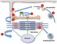
          Centronuclear myopathies (CNMs) are a genetically heterogeneous group of inherited neuromuscular disorders characterized by clinical features of a congenital myopathy and abundant central nuclei as the most prominent histopathological feature. The most common forms of congenital myopathies with central nuclei have been attributed to X-linked recessive mutations in the MTM1 gene encoding myotubularin (“X-linked myotubular myopathy”), autosomal-dominant mutations in the DNM2 gene encoding dynamin-2 and the BIN1 gene encoding amphiphysin-2 (also named bridging integrator-1, BIN1, or SH3P9), and autosomal-recessive mutations in BIN1, the RYR1 gene encoding the skeletal muscle ryanodine receptor, and the TTN gene encoding titin. Models to study and rescue the affected cellular pathways are now available in yeast, C. elegans, drosophila, zebrafish, mouse, and dog. Defects in membrane trafficking have emerged as a key pathogenic mechanisms, with aberrant T-tubule formation, abnormalities of triadic assembly, and disturbance of the excitation–contraction machinery the main downstream effects studied to date. Abnormal autophagy has recently been recognized as another important collateral of defective membrane trafficking in different genetic forms of CNM, suggesting an intriguing link to primary disorders of defective autophagy with overlapping histopathological features. The following review will provide an overview of clinical, histopathological, and genetic aspects of the CNMs in the context of the key pathogenic mechanism, outline unresolved questions, and indicate promising future lines of enquiry.

          Related collections

          Most cited references113

          • Record: found
          • Abstract: found
          • Article: not found

          BAR domains as sensors of membrane curvature: the amphiphysin BAR structure.

          The BAR (Bin/amphiphysin/Rvs) domain is the most conserved feature in amphiphysins from yeast to human and is also found in endophilins and nadrins. We solved the structure of the Drosophila amphiphysin BAR domain. It is a crescent-shaped dimer that binds preferentially to highly curved negatively charged membranes. With its N-terminal amphipathic helix and BAR domain (N-BAR), amphiphysin can drive membrane curvature in vitro and in vivo. The structure is similar to that of arfaptin2, which we find also binds and tubulates membranes. From this, we predict that BAR domains are in many protein families, including sorting nexins, centaurins, and oligophrenins. The universal and minimal BAR domain is a dimerization, membrane-binding, and curvature-sensing module.
            Bookmark
            • Record: found
            • Abstract: found
            • Article: not found

            The Beclin 1-VPS34 complex--at the crossroads of autophagy and beyond.

            An increasing body of research on autophagy provides overwhelming evidence for its connection to diverse biological functions and human diseases. Beclin 1, the first mammalian autophagy protein to be described, appears to act as a nexus point between autophagy, endosomal, and perhaps also cell death pathways. Beclin 1 performs these roles as part of a core complex that contains vacuolar sorting protein 34 (VPS34), a class III phosphatidylinositol-3 kinase. The precise mechanism of Beclin 1-mediated regulation of these cellular functions is unclear, but substantial progress has recently been made in identifying new players and their functions in Beclin 1-VSP34 complexes. Here we review emerging studies that are beginning to unveil the physiological functions of Beclin 1-VPS34 in the central control of autophagic activity and other trafficking events through the formation of distinct Beclin 1-VPS34 protein complexes. (c) 2010 Elsevier Ltd. All rights reserved.
              Bookmark
              • Record: found
              • Abstract: found
              • Article: not found

              The regulation and function of Class III PI3Ks: novel roles for Vps34.

              The Class III PI3K (phosphoinositide 3-kinase), Vps34 (vacuolar protein sorting 34), was first described as a component of the vacuolar sorting system in Saccharomyces cerevisiae and is the sole PI3K in yeast. The homologue in mammalian cells, hVps34, has been studied extensively in the context of endocytic sorting. However, hVps34 also plays an important role in the ability of cells to respond to changes in nutrient conditions. Recent studies have shown that mammalian hVps34 is required for the activation of the mTOR (mammalian target of rapamycin)/S6K1 (S6 kinase 1) pathway, which regulates protein synthesis in response to nutrient availability. In both yeast and mammalian cells, Class III PI3Ks are also required for the induction of autophagy during nutrient deprivation. Finally, mammalian hVps34 is itself regulated by nutrients. Thus Class III PI3Ks are implicated in the regulation of both autophagy and, through the mTOR pathway, protein synthesis, and thus contribute to the integration of cellular responses to changing nutritional status.
                Bookmark

                Author and article information

                Contributors
                Journal
                Front Aging Neurosci
                Front Aging Neurosci
                Front. Aging Neurosci.
                Frontiers in Aging Neuroscience
                Frontiers Media S.A.
                1663-4365
                06 November 2014
                19 December 2014
                2014
                : 6
                : 339
                Affiliations
                [1] 1Neuromuscular Service, Department of Paediatric Neurology, Evelina Children’s Hospital, St Thomas’ Hospital , London, UK
                [2] 2Department of Basic and Clinical Neuroscience, Institute of Psychiatry, Psychology and Neuroscience (IoPPN), King’s College London , London, UK
                [3] 3Randall Division of Cell and Molecular Biophysics and Cardiovascular Division, King’s College London BHF Centre of Research Excellence , London, UK
                Author notes

                Edited by: Luciano Merlini, Istituto Ortopedico Rizzoli IRCCS, Italy

                Reviewed by: Jocelyn Laporte, INSERM, France; Denise Alessandra Cassandrini, IRCCS Fondazione Stella Maris, Italy

                *Correspondence: Heinz Jungbluth, Children’s Neuroscience Centre, St Thomas’ Hospital, Floor 01 – Staircase D South Wing, London SE1 7EH, UK e-mail: heinz.jungbluth@ 123456gstt.nhs.uk

                This article was submitted to the journal Frontiers in Aging Neuroscience.

                Article
                10.3389/fnagi.2014.00339
                4271577
                25566070
                a67046f1-8f5f-49a9-8aca-8881863792f8
                Copyright © 2014 Jungbluth and Gautel.

                This is an open-access article distributed under the terms of the Creative Commons Attribution License (CC BY). The use, distribution or reproduction in other forums is permitted, provided the original author(s) or licensor are credited and that the original publication in this journal is cited, in accordance with accepted academic practice. No use, distribution or reproduction is permitted which does not comply with these terms.

                History
                : 22 October 2014
                : 02 December 2014
                Page count
                Figures: 1, Tables: 1, Equations: 0, References: 132, Pages: 11, Words: 10238
                Categories
                Neuroscience
                Review Article

                Neurosciences
                centronuclear myopathy,myotubular myopathy,mtm1 myotubularin gene,dnm2 dynamin-2 gene,bin1 bridging integrator-1/amphiphysin-2 gene,ryr1 ryanodine receptor-1 gene,ttn titin gene,autophagy

                Comments

                Comment on this article