23
views
0
recommends
+1 Recommend
0 collections
    0
    shares
      • Record: found
      • Abstract: found
      • Article: not found

      Pigmented eccrine poroma: dermoscopic and confocal features

      research-article

      Read this article at

      ScienceOpenPublisherPMC
      Bookmark
          There is no author summary for this article yet. Authors can add summaries to their articles on ScienceOpen to make them more accessible to a non-specialist audience.

          Abstract

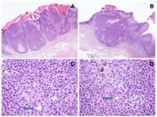
          Eccrine poroma is a rare benign adnexal tumor of epithelial cells originating from the terminal ductal portion of the sweat glands that is typically located on palms and soles, although other cutaneous sites can be affected [ 1]. It is usually nonpigmented even if there is a pigmented variant that corresponds to 17% of cases and it is usually underdiagnosed, since it is mistakenly confused with other pigmented tumors [ 2, 3]. Dermoscopy and reflectance confocal microscopy (RCM) may assist in the correct diagnosis of this tumor.

          Herein, we report one case of pigmented eccrine poroma (PEP) that simulated clinically a cutaneous melanoma or a basal cell carcinoma. Dermoscopy and RCM excluded the possibilities of those two diagnoses; the overall confocal findings were suggestive for a benign epithelial tumor. Histology was fundamental to diagnose this lesion as a pigmented eccrine poroma. Even if the diagnosis of eccrine poroma remains histopathological still, as in this case report, noninvasive tools such as dermoscopy and RCM examinations can be of help to rule out the diagnosis of melanoma. Larger studies on this rare pigmented variant of eccrine poroma could shed new light on the identification of specific diagnostic dermoscopic and confocal features.

          Related collections

          Most cited references11

          • Record: found
          • Abstract: found
          • Article: not found

          Reflectance confocal microscopy as a second-level examination in skin oncology improves diagnostic accuracy and saves unnecessary excisions: a longitudinal prospective study.

          Dermatoscopy increases both the sensitivity and specificity of melanoma diagnosis. Reflectance confocal microscopy (RCM) is a noninvasive technique that complements dermatoscopy in the evaluation of equivocal lesions at cellular resolution. To determine prospectively the potential impact of confocal microscopy when implemented in a routine melanoma diagnosis workflow. Patients referred to a single melanoma clinic were consecutively enrolled. At dermatoscopy, patients were referred to one of the following pathways: (i) no further examination or (ii) RCM examination. On examination atypical lesion(s) were referred for either (a) RCM documentation (lesions with consistent suspicious clinical/dermatoscopic criteria, already qualified and scheduled for surgical excision) or (b) RCM consultation for equivocal lesions, where RCM diagnosis would determine lesion definite outcome (excision or digital follow-up). Reflectance confocal microscopy examination was performed for 41% of 1005 patients enrolled. In two-thirds of these cases RCM influenced the lesion outcome. The systematic application of RCM for equivocal lesions saved over 50% of benign lesions from unnecessary excision. The number needed to excise a melanoma was 6·8 with RCM examination, compared with a hypothetical 14·6 without RCM evaluation. Reflectance confocal microscopy as a second-level examination to dermatoscopy proved to be highly accurate in diagnosis and reduced the number of unnecessary excisions. Improved accuracy, considering that RCM enabled the detection of the six melanomas (2%) in the group of 308 lesions eligible for follow-up, also minimizes the risk of referring a melanoma to digital dermatoscopy monitoring, and potentially losing the patient to follow-up. © 2014 British Association of Dermatologists.
            Bookmark
            • Record: found
            • Abstract: found
            • Article: not found

            Classifying distinct basal cell carcinoma subtype by means of dermatoscopy and reflectance confocal microscopy.

            The current guidelines for the management of basal cell carcinoma (BCC) suggest a different therapeutic approach according to histopathologic subtype. Although dermatoscopic and confocal criteria of BCC have been investigated, no specific studies were performed to evaluate the distinct reflectance confocal microscopy (RCM) aspects of BCC subtypes.
              Bookmark
              • Record: found
              • Abstract: not found
              • Article: not found

              Eccrine poroma; tumors exhibiting features of the epidermal sweat duct unit.

                Bookmark

                Author and article information

                Journal
                Dermatol Pract Concept
                Dermatol Pract Concept
                DP
                Dermatology Practical & Conceptual
                Derm101.com
                2160-9381
                July 2016
                31 July 2016
                : 6
                : 3
                : 59-62
                Affiliations
                [1 ]Dermatology Unit, University of Modena and Reggio Emilia, Italy
                [2 ]Pathology Unit, Arcispedale S. Maria Nuova, IRCCS, Reggio Emilia, Italy
                [3 ]Dermatology and Skin Cancer Unit, Arcispedale S. Maria Nuova, IRCCS, Reggio Emilia, Italy
                [4 ]First Department of Dermatology, Aristotle University, Thessaloniki, Greece
                [5 ]Dermatology Unit, Second University of Naples, Naples, Italy
                Author notes
                Corresponding author: Caterina Longo, MD, PhD, Dermatology and Skin Cancer Unit, Arcispedale Santa Maria Nuova-IRCCS, Viale Risorgimento, 80, 42100 Reggio Emilia, Italy. Tel. +390522295612; Fax. +390594224271. Email: longo.caterina@ 123456gmail.com
                Article
                dp0603a12
                10.5826/dpc.0603a12
                5006555
                27648386
                a5b6a9b8-8777-4499-a7a7-2ad9d12d50fd
                Copyright: ©2016 Bombonato et al.

                This is an open-access article distributed under the terms of the Creative Commons Attribution License, which permits unrestricted use, distribution, and reproduction in any medium, provided the original author and source are credited.

                History
                : 23 April 2016
                : 27 May 2016
                Categories
                Observation

                Comments

                Comment on this article