2
views
0
recommends
+1 Recommend
0 collections
    0
    shares
      • Record: found
      • Abstract: found
      • Article: found
      Is Open Access

      The Beneficial Effect of Mitochondrial Transfer Therapy in 5XFAD Mice via Liver–Serum–Brain Response

      Read this article at

      Bookmark
          There is no author summary for this article yet. Authors can add summaries to their articles on ScienceOpen to make them more accessible to a non-specialist audience.

          Abstract

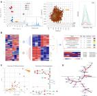
          We recently reported the benefit of the IV transferring of active exogenous mitochondria in a short-term pharmacological AD (Alzheimer’s disease) model. We have now explored the efficacy of mitochondrial transfer in 5XFAD transgenic mice, aiming to explore the underlying mechanism by which the IV-injected mitochondria affect the diseased brain. Mitochondrial transfer in 5XFAD ameliorated cognitive impairment, amyloid burden, and mitochondrial dysfunction. Exogenously injected mitochondria were detected in the liver but not in the brain. We detected alterations in brain proteome, implicating synapse-related processes, ubiquitination/proteasome-related processes, phagocytosis, and mitochondria-related factors, which may lead to the amelioration of disease. These changes were accompanied by proteome/metabolome alterations in the liver, including pathways of glucose, glutathione, amino acids, biogenic amines, and sphingolipids. Altered liver metabolites were also detected in the serum of the treated mice, particularly metabolites that are known to affect neurodegenerative processes, such as carnosine, putrescine, C24:1-OH sphingomyelin, and amino acids, which serve as neurotransmitters or their precursors. Our results suggest that the beneficial effect of mitochondrial transfer in the 5XFAD mice is mediated by metabolic signaling from the liver via the serum to the brain, where it induces protective effects. The high efficacy of the mitochondrial transfer may offer a novel AD therapy.

          Related collections

          Most cited references73

          • Record: found
          • Abstract: found
          • Article: found
          Is Open Access

          MetaboAnalyst 5.0: narrowing the gap between raw spectra and functional insights

          Since its first release over a decade ago, the MetaboAnalyst web-based platform has become widely used for comprehensive metabolomics data analysis and interpretation. Here we introduce MetaboAnalyst version 5.0, aiming to narrow the gap from raw data to functional insights for global metabolomics based on high-resolution mass spectrometry (HRMS). Three modules have been developed to help achieve this goal, including: (i) a LC–MS Spectra Processing module which offers an easy-to-use pipeline that can perform automated parameter optimization and resumable analysis to significantly lower the barriers to LC-MS1 spectra processing; (ii) a Functional Analysis module which expands the previous MS Peaks to Pathways module to allow users to intuitively select any peak groups of interest and evaluate their enrichment of potential functions as defined by metabolic pathways and metabolite sets; (iii) a Functional Meta-Analysis module to combine multiple global metabolomics datasets obtained under complementary conditions or from similar studies to arrive at comprehensive functional insights. There are many other new functions including weighted joint-pathway analysis, data-driven network analysis, batch effect correction, merging technical replicates, improved compound name matching, etc. The web interface, graphics and underlying codebase have also been refactored to improve performance and user experience. At the end of an analysis session, users can now easily switch to other compatible modules for a more streamlined data analysis. MetaboAnalyst 5.0 is freely available at https://www.metaboanalyst.ca . Graphical Abstract From raw data to statistical and functional insights using MetaboAnalyst 5.0.
            Bookmark
            • Record: found
            • Abstract: found
            • Article: not found

            Intraneuronal beta-amyloid aggregates, neurodegeneration, and neuron loss in transgenic mice with five familial Alzheimer's disease mutations: potential factors in amyloid plaque formation.

            Mutations in the genes for amyloid precursor protein (APP) and presenilins (PS1, PS2) increase production of beta-amyloid 42 (Abeta42) and cause familial Alzheimer's disease (FAD). Transgenic mice that express FAD mutant APP and PS1 overproduce Abeta42 and exhibit amyloid plaque pathology similar to that found in AD, but most transgenic models develop plaques slowly. To accelerate plaque development and investigate the effects of very high cerebral Abeta42 levels, we generated APP/PS1 double transgenic mice that coexpress five FAD mutations (5XFAD mice) and additively increase Abeta42 production. 5XFAD mice generate Abeta42 almost exclusively and rapidly accumulate massive cerebral Abeta42 levels. Amyloid deposition (and gliosis) begins at 2 months and reaches a very large burden, especially in subiculum and deep cortical layers. Intraneuronal Abeta42 accumulates in 5XFAD brain starting at 1.5 months of age (before plaques form), is aggregated (as determined by thioflavin S staining), and occurs within neuron soma and neurites. Some amyloid deposits originate within morphologically abnormal neuron soma that contain intraneuronal Abeta. Synaptic markers synaptophysin, syntaxin, and postsynaptic density-95 decrease with age in 5XFAD brain, and large pyramidal neurons in cortical layer 5 and subiculum are lost. In addition, levels of the activation subunit of cyclin-dependent kinase 5, p25, are elevated significantly at 9 months in 5XFAD brain, although an upward trend is observed by 3 months of age, before significant neurodegeneration or neuron loss. Finally, 5XFAD mice have impaired memory in the Y-maze. Thus, 5XFAD mice rapidly recapitulate major features of AD amyloid pathology and may be useful models of intraneuronal Abeta42-induced neurodegeneration and amyloid plaque formation.
              Bookmark
              • Record: found
              • Abstract: found
              • Article: found
              Is Open Access

              The PI3K/AKT pathway in obesity and type 2 diabetes

              Obesity and type 2 diabetes mellitus are complicated metabolic diseases that affect multiple organs and are characterized by hyperglycaemia. Currently, stable and effective treatments for obesity and type 2 diabetes mellitus are not available. Therefore, the mechanisms leading to obesity and diabetes and more effective ways to treat obesity and diabetes should be identified. Based on accumulated evidences, the PI3K/AKT signalling pathway is required for normal metabolism due to its characteristics, and its imbalance leads to the development of obesity and type 2 diabetes mellitus. This review focuses on the role of PI3K/AKT signalling in the skeletal muscle, adipose tissue, liver, brain and pancreas, and discusses how this signalling pathway affects the development of the aforementioned diseases. We also summarize evidences for recently identified therapeutic targets of the PI3K/AKT pathway as treatments for obesity and type 2 diabetes mellitus. PI3K/AKT pathway damaged in various tissues of the body leads to obesity and type 2 diabetes as the result of insulin resistance, and in turn, insulin resistance exacerbates the PI3K/AKT pathway, forming a vicious circle.
                Bookmark

                Author and article information

                Contributors
                (View ORCID Profile)
                (View ORCID Profile)
                (View ORCID Profile)
                (View ORCID Profile)
                (View ORCID Profile)
                (View ORCID Profile)
                Journal
                CELLC6
                Cells
                Cells
                MDPI AG
                2073-4409
                April 2023
                March 24 2023
                : 12
                : 7
                : 1006
                Article
                10.3390/cells12071006
                a559569c-3d00-4512-a230-7003e64b0074
                © 2023

                https://creativecommons.org/licenses/by/4.0/

                History

                Comments

                Comment on this article