3
views
0
recommends
+1 Recommend
0 collections
    0
    shares
      • Record: found
      • Abstract: found
      • Article: found
      Is Open Access

      Oncogenic Gain of Function in Glioblastoma Is Linked to Mutant p53 Amyloid Oligomers

      research-article

      Read this article at
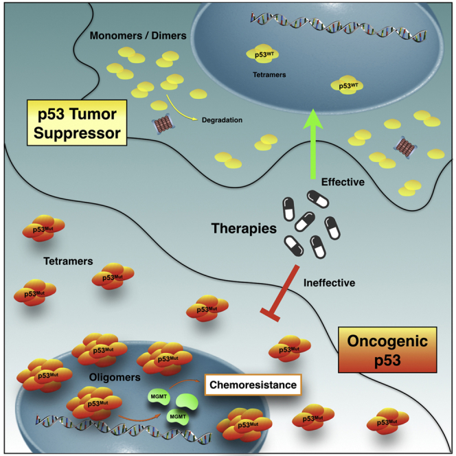
      Bookmark
          There is no author summary for this article yet. Authors can add summaries to their articles on ScienceOpen to make them more accessible to a non-specialist audience.

          Summary

          Tumor-associated p53 mutations endow cells with malignant phenotypes, including chemoresistance. Amyloid-like oligomers of mutant p53 transform this tumor suppressor into an oncogene. However, the composition and distribution of mutant p53 oligomers are unknown and the mechanism involved in the conversion is sparse. Here, we report accumulation of a p53 mutant within amyloid-like p53 oligomers in glioblastoma-derived cells presenting a chemoresistant gain-of-function phenotype. Statistical analysis from fluorescence fluctuation spectroscopy, pressure-induced measurements, and thioflavin T kinetics demonstrates the distribution of oligomers larger than the active tetrameric form of p53 in the nuclei of living cells and the destabilization of native-drifted p53 species that become amyloid. Collectively, these results provide insights into the role of amyloid-like mutant p53 oligomers in the chemoresistance phenotype of malignant and invasive brain tumors and shed light on therapeutic options to avert cancer.

          Graphical Abstract

          Highlights

          • Amyloid oligomers transform p53 tumor suppressor into an oncogene

          • Amyloid-like mutant p53 oligomers occur in chemoresistant glioblastoma cells

          • p53 oligomer larger than tetramers is detected in the nuclei of living cells

          • Gain-of-function p53 phenotypes is attributed to p53 amyloid oligomers

          Abstract

          Structural Biology; Protein Structure Aspects; Biophysics; Cancer

          Related collections

          Most cited references57

          • Record: found
          • Abstract: found
          • Article: not found

          DNA damage-induced phosphorylation of p53 alleviates inhibition by MDM2.

          DNA-damaging agents signal to p53 through as yet unidentified posttranscriptional mechanisms. Here we show that phosphorylation of human p53 at serine 15 occurs after DNA damage and that this leads to reduced interaction of p53 with its negative regulator, the oncoprotein MDM2, in vivo and in vitro. Furthermore, using purified DNA-dependent protein kinase (DNA-PK), we demonstrate that phosphorylation of p53 at serines 15 and 37 impairs the ability of MDM2 to inhibit p53-dependent transactivation. We present evidence that these effects are most likely due to a conformational change induced upon phosphorylation of p53. Our studies provide a plausible mechanism by which the induction of p53 can be modulated by DNA-PK (or other protein kinases with similar specificity) in response to DNA damage.
            Bookmark
            • Record: found
            • Abstract: found
            • Article: found

            Genetic pathways to primary and secondary glioblastoma.

            Glioblastoma is the most frequent and most malignant human brain tumor. The prognosis remains very poor, with most patients dying within 1 year after diagnosis. Primary and secondary glioblastoma constitute distinct disease subtypes, affecting patients of different age and developing through different genetic pathways. The majority of cases (>90%) are primary glioblastomas that develop rapidly de novo, without clinical or histological evidence of a less malignant precursor lesion. They affect mainly the elderly and are genetically characterized by loss of heterozygosity 10q (70% of cases), EGFR amplification (36%), p16(INK4a) deletion (31%), and PTEN mutations (25%). Secondary glioblastomas develop through progression from low-grade diffuse astrocytoma or anaplastic astrocytoma and manifest in younger patients. In the pathway to secondary glioblastoma, TP53 mutations are the most frequent and earliest detectable genetic alteration, already present in 60% of precursor low-grade astrocytomas. The mutation pattern is characterized by frequent G:C-->A:T mutations at CpG sites. During progression to glioblastoma, additional mutations accumulate, including loss of heterozygosity 10q25-qter ( approximately 70%), which is the most frequent genetic alteration in both primary and secondary glioblastomas. Primary and secondary glioblastomas also differ significantly in their pattern of promoter methylation and in expression profiles at RNA and protein levels. This has significant implications, particularly for the development of novel, targeted therapies, as discussed in this review.
              Bookmark
              • Record: found
              • Abstract: found
              • Article: not found

              Rescue of early embryonic lethality in mdm2-deficient mice by deletion of p53.

              The gene p53 encodes a transcriptional activator of genes involved in growth arrest, DNA repair and apoptosis. Loss of p53 function contributes to tumour development in vivo. The transcriptional activation function of p53 is inactivated by interaction with the mdm2 gene product. Amplification of mdm2 has been observed in 36% of human sarcomas, indicating that it may represent an alternative mechanism of preventing p53 function in tumour development. To study mdm2 function in vivo, we generated an mdm2 null allele by homologous recombination. Mdm2 null mice are not viable, and further analysis revealed embryonic lethality around implantation. To examine the importance of the interaction of MDM2 with p53 in vivo, we crossed mice heterozygous for mdm2 and p53 and obtained progeny homozygous for both p53 and mdm2 null alleles. Rescue of the mdm2-/- lethality in a p53 null background suggests that a critical in vivo function of MDM2 is the negative regulation of p53 activity.
                Bookmark

                Author and article information

                Contributors
                Journal
                iScience
                iScience
                iScience
                Elsevier
                2589-0042
                08 January 2020
                21 February 2020
                08 January 2020
                : 23
                : 2
                : 100820
                Affiliations
                [1 ]Institute of Medical Biochemistry Leopoldo de Meis, National Institute of Science and Technology for Structural Biology and Bioimaging, National Center of Nuclear Magnetic Resonance Jiri Jonas, Federal University of Rio de Janeiro, Rio de Janeiro, Rio de Janeiro 21941-901, Brazil
                [2 ]Universidade Federal do ABC, Centro de Ciências Naturais e Humanas. Av. dos Estados, 5001 Sta. Terezinha, Santo André, São Paulo 21941-590, Brazil
                [3 ]Laboratório de Biomedicina do Cérebro, Instituto Estadual do Cérebro Paulo Niemeyer (IECPN), Secretaria de Estado de Saúde, Rio de Janeiro, Brazil
                [4 ]Instituto de Ciências Biomédicas, Universidade Federal do Rio de Janeiro, Rio de Janeiro, Brazil
                [5 ]Laboratory for Fluorescence Dynamics, Biomedical Engineering Department, University of California, Irvine, CA 92697-2717, USA
                [6 ]Department of Biochemistry and Molecular Genetics, University of Virginia, Charlottesville, VA 22908-0733, USA
                Author notes
                []Corresponding author jerson@ 123456bioqmed.ufrj.br
                [∗∗ ]Corresponding author gaugusto@ 123456bioqmed.ufrj.br
                [7]

                Lead Contact

                Article
                S2589-0042(20)30003-1 100820
                10.1016/j.isci.2020.100820
                6976948
                31981923
                a53f39ba-5c0e-4a3f-94ca-99a6cdf9942b
                © 2020 The Author(s)

                This is an open access article under the CC BY license (http://creativecommons.org/licenses/by/4.0/).

                History
                : 21 August 2019
                : 20 November 2019
                : 2 January 2020
                Categories
                Article

                structural biology,protein structure aspects,biophysics,cancer

                Comments

                Comment on this article