0
views
0
recommends
+1 Recommend
0 collections
    0
    shares
      • Record: found
      • Abstract: found
      • Article: found
      Is Open Access

      Plasma Neurofilament Light Chain and Clinical Diagnosis in Frontotemporal Dementia Syndromes

      research-article

      Read this article at

      Bookmark
          There is no author summary for this article yet. Authors can add summaries to their articles on ScienceOpen to make them more accessible to a non-specialist audience.

          Abstract

          Background:

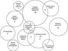
          Frontotemporal dementia (FTD) syndromes, mimics, phenocopy (phFTD), and slowly progressive behavioral variant FTD (bvFTD) can be difficult to distinguish clinically. Biomarkers such as neurofilament light chain (NfL) may be helpful.

          Objective:

          To study plasma NfL levels in people with FTD syndromes and determine if plasma NfL can distinguish between FTD syndromes and phFTD.

          Methods:

          Plasma NfL levels were estimated using both Simoa ® Quanterix HD-X™ and SR-X™ machines grouped via final diagnosis after investigation and review.

          Results:

          Fifty participants were studied: bvFTD = 20, semantic variant FTD (svFTD) = 11, non-fluent variant FTD (nfvFTD) = 9, FTD with motor neuron disease (MND) = 4, phFTD = 2, slow progressors = 3, FTD mimic = 1, mean age 67.2 (SD 8.4) years. NfL levels were significantly higher in the FTD group compared to phenocopy group ( p = 0.003). Median NfL (IQR) pg/mL was comparable in the FTD syndromes: bvFTD 41.10 (50.72), svFTD 44.38 (16.61), and nfvFTD 42.61 (22.93), highest in FTD with MND 79.67 (45.32) and lowest in both phFTD 13.99 (0.79) and slow progressors 17.97 (3.62).

          Conclusion:

          Plasma NfL appears to differentiate FTD syndromes and mimics. However, a lower NfL may predict a slower, but not necessarily absence of neurodegeneration, and therefore appears limited in distinguishing slow progressors from FTD phenocopies. Larger numbers of patients from all clinical groups are required to strengthen diagnostic utility.

          Related collections

          Most cited references49

          • Record: found
          • Abstract: found
          • Article: found

          Classification of primary progressive aphasia and its variants

          This article provides a classification of primary progressive aphasia (PPA) and its 3 main variants to improve the uniformity of case reporting and the reliability of research results. Criteria for the 3 variants of PPA--nonfluent/agrammatic, semantic, and logopenic--were developed by an international group of PPA investigators who convened on 3 occasions to operationalize earlier published clinical descriptions for PPA subtypes. Patients are first diagnosed with PPA and are then divided into clinical variants based on specific speech and language features characteristic of each subtype. Classification can then be further specified as "imaging-supported" if the expected pattern of atrophy is found and "with definite pathology" if pathologic or genetic data are available. The working recommendations are presented in lists of features, and suggested assessment tasks are also provided. These recommendations have been widely agreed upon by a large group of experts and should be used to ensure consistency of PPA classification in future studies. Future collaborations will collect prospective data to identify relationships between each of these syndromes and specific biomarkers for a more detailed understanding of clinicopathologic correlations.
            Bookmark
            • Record: found
            • Abstract: found
            • Article: found

            Neurofilament light chain as a biomarker in neurological disorders

            In the management of neurological diseases, the identification and quantification of axonal damage could allow for the improvement of diagnostic accuracy and prognostic assessment. Neurofilament light chain (NfL) is a neuronal cytoplasmic protein highly expressed in large calibre myelinated axons. Its levels increase in cerebrospinal fluid (CSF) and blood proportionally to the degree of axonal damage in a variety of neurological disorders, including inflammatory, neurodegenerative, traumatic and cerebrovascular diseases. New immunoassays able to detect biomarkers at ultralow levels have allowed for the measurement of NfL in blood, thus making it possible to easily and repeatedly measure NfL for monitoring diseases’ courses. Evidence that both CSF and blood NfL may serve as diagnostic, prognostic and monitoring biomarkers in neurological diseases is progressively increasing, and NfL is one of the most promising biomarkers to be used in clinical and research setting in the next future. Here we review the most important results on CSF and blood NfL and we discuss its potential applications and future directions.
              Bookmark
              • Record: found
              • Abstract: found
              • Article: not found

              Comparison of three analytical platforms for quantification of the neurofilament light chain in blood samples: ELISA, electrochemiluminescence immunoassay and Simoa.

              Neuronal damage is the morphological substrate of persisting neurological disability. Neurofilaments (Nf) are specific cytoskeletal proteins of neurons and their quantification has shown encouraging results as a biomarker for axonal injury.
                Bookmark

                Author and article information

                Journal
                J Alzheimers Dis
                J Alzheimers Dis
                JAD
                Journal of Alzheimer's Disease
                IOS Press (Nieuwe Hemweg 6B, 1013 BG Amsterdam, The Netherlands )
                1387-2877
                1875-8908
                17 August 2022
                11 October 2022
                2022
                : 89
                : 4
                : 1221-1231
                Affiliations
                [a ]Eastern Cognitive Disorders Clinic, Eastern Health, Box Hill, VIC, Australia
                [b ] Eastern Clinical Research Unit, Eastern Health Clinical School, Faculty of Medicine, Nursing and Health Sciences, Monash University , Melbourne, VIC, Australia
                [c ] Royal Melbourne Hospital, Department of Neurology, Parkville, VIC, Australia
                [d ] Florey Institute of Neuroscience and Mental Health , Parkville, VIC, Australia
                [e ] Neuropsychiatry and Melbourne Neuropsychiatry Centre, Royal Melbourne Hospital and University of Melbourne , Parkville, VIC, Australia
                [f ]Alfred Health, Department of Neurology, Prahran, VIC, Australia
                [g ]Calvary Health Care Bethlehem, Parkdale, VIC, Australia
                Author notes
                [* ]Correspondence to: Suyi Ooi, Eastern Health, Department of Neurosciences, 5 Arnold Street, Box Hill, Victoria 3128, Australia. E-mail: suyi.ooi@ 123456easternhealth.org.au .
                Article
                JAD220272
                10.3233/JAD-220272
                9661326
                35988220
                a4bbdd6d-7e77-4ed0-a788-ceebc33c5397
                © 2022 – The authors. Published by IOS Press

                This is an open access article distributed under the terms of the Creative Commons Attribution Non-Commercial (CC BY-NC 4.0) License, which permits unrestricted non-commercial use, distribution, and reproduction in any medium, provided the original work is properly cited.

                History
                : 25 July 2022
                Categories
                Research Article

                biomarker,frontotemporal dementia,neurofilament light,phenocopy,primary progressive aphasia

                Comments

                Comment on this article