11
views
0
recommends
+1 Recommend
0 collections
    0
    shares
      • Record: found
      • Abstract: found
      • Article: found
      Is Open Access

      Dihydroceramide accumulation mediates cytotoxic autophagy of cancer cells via autolysosome destabilization

      research-article

      Read this article at

      Bookmark
          There is no author summary for this article yet. Authors can add summaries to their articles on ScienceOpen to make them more accessible to a non-specialist audience.

          ABSTRACT

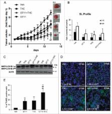
          Autophagy is considered primarily a cell survival process, although it can also lead to cell death. However, the factors that dictate the shift between these 2 opposite outcomes remain largely unknown. In this work, we used Δ 9-tetrahydrocannabinol (THC, the main active component of marijuana, a compound that triggers autophagy-mediated cancer cell death) and nutrient deprivation (an autophagic stimulus that triggers cytoprotective autophagy) to investigate the precise molecular mechanisms responsible for the activation of cytotoxic autophagy in cancer cells. By using a wide array of experimental approaches we show that THC (but not nutrient deprivation) increases the dihydroceramide:ceramide ratio in the endoplasmic reticulum of glioma cells, and this alteration is directed to autophagosomes and autolysosomes to promote lysosomal membrane permeabilization, cathepsin release and the subsequent activation of apoptotic cell death. These findings pave the way to clarify the regulatory mechanisms that determine the selective activation of autophagy-mediated cancer cell death.

          Related collections

          Most cited references45

          • Record: found
          • Abstract: found
          • Article: not found

          Molecular characterization of a peripheral receptor for cannabinoids.

          The major active ingredient of marijuana, delta 9-tetrahydrocannabinol (delta 9-THC), has been used as a psychoactive agent for thousands of years. Marijuana, and delta 9-THC, also exert a wide range of other effects including analgesia, anti-inflammation, immunosuppression, anticonvulsion, alleviation of intraocular pressure in glaucoma, and attenuation of vomiting. The clinical application of cannabinoids has, however, been limited by their psychoactive effects, and this has led to interest in the biochemical bases of their action. Progress stemmed initially from the synthesis of potent derivatives of delta 9-THC, and more recently from the cloning of a gene encoding a G-protein-coupled receptor for cannabinoids. This receptor is expressed in the brain but not in the periphery, except for a low level in testes. It has been proposed that the nonpsychoactive effects of cannabinoids are either mediated centrally or through direct interaction with other, non-receptor proteins. Here we report the cloning of a receptor for cannabinoids that is not expressed in the brain but rather in macrophages in the marginal zone of spleen.
            Bookmark
            • Record: found
            • Abstract: found
            • Article: not found

            Structure of a cannabinoid receptor and functional expression of the cloned cDNA.

            Marijuana and many of its constituent cannabinoids influence the central nervous system (CNS) in a complex and dose-dependent manner. Although CNS depression and analgesia are well documented effects of the cannabinoids, the mechanisms responsible for these and other cannabinoid-induced effects are not so far known. The hydrophobic nature of these substances has suggested that cannabinoids resemble anaesthetic agents in their action, that is, they nonspecifically disrupt cellular membranes. Recent evidence, however, has supported a mechanism involving a G protein-coupled receptor found in brain and neural cell lines, and which inhibits adenylate cyclase activity in a dose-dependent, stereoselective and pertussis toxin-sensitive manner. Also, the receptor is more responsive to psychoactive cannabinoids than to non-psychoactive cannabinoids. Here we report the cloning and expression of a complementary DNA that encodes a G protein-coupled receptor with all of these properties. Its messenger RNA is found in cell lines and regions of the brain that have cannabinoid receptors. These findings suggest that this protein is involved in cannabinoid-induced CNS effects (including alterations in mood and cognition) experienced by users of marijuana.
              Bookmark
              • Record: found
              • Abstract: found
              • Article: not found

              Guidelines for the use and interpretation of assays for monitoring autophagy.

              In 2008 we published the first set of guidelines for standardizing research in autophagy. Since then, research on this topic has continued to accelerate, and many new scientists have entered the field. Our knowledge base and relevant new technologies have also been expanding. Accordingly, it is important to update these guidelines for monitoring autophagy in different organisms. Various reviews have described the range of assays that have been used for this purpose. Nevertheless, there continues to be confusion regarding acceptable methods to measure autophagy, especially in multicellular eukaryotes. A key point that needs to be emphasized is that there is a difference between measurements that monitor the numbers or volume of autophagic elements (e.g., autophagosomes or autolysosomes) at any stage of the autophagic process vs. those that measure flux through the autophagy pathway (i.e., the complete process); thus, a block in macroautophagy that results in autophagosome accumulation needs to be differentiated from stimuli that result in increased autophagic activity, defined as increased autophagy induction coupled with increased delivery to, and degradation within, lysosomes (in most higher eukaryotes and some protists such as Dictyostelium) or the vacuole (in plants and fungi). In other words, it is especially important that investigators new to the field understand that the appearance of more autophagosomes does not necessarily equate with more autophagy. In fact, in many cases, autophagosomes accumulate because of a block in trafficking to lysosomes without a concomitant change in autophagosome biogenesis, whereas an increase in autolysosomes may reflect a reduction in degradative activity. Here, we present a set of guidelines for the selection and interpretation of methods for use by investigators who aim to examine macroautophagy and related processes, as well as for reviewers who need to provide realistic and reasonable critiques of papers that are focused on these processes. These guidelines are not meant to be a formulaic set of rules, because the appropriate assays depend in part on the question being asked and the system being used. In addition, we emphasize that no individual assay is guaranteed to be the most appropriate one in every situation, and we strongly recommend the use of multiple assays to monitor autophagy. In these guidelines, we consider these various methods of assessing autophagy and what information can, or cannot, be obtained from them. Finally, by discussing the merits and limits of particular autophagy assays, we hope to encourage technical innovation in the field.
                Bookmark

                Author and article information

                Journal
                Autophagy
                Autophagy
                KAUP
                kaup20
                Autophagy
                Taylor & Francis
                1554-8627
                1554-8635
                2016
                16 September 2016
                16 September 2016
                : 12
                : 11
                : 2213-2229
                Affiliations
                [a ]Department of Biochemistry and Molecular Biology I, School of Biology, Complutense University , Madrid, Spain
                [b ]Instituto de Investigaciones Sanitarias San Carlos (IdISSC) , Madrid, Spain
                [c ]Research Unit on BioActive Molecules (RUBAM), Departments of Biomedicinal Chemistry, Institute for Advanced Chemistry of Catalonia (IQAC-CSIC) , Barcelona, Spain
                [d ]Biofisika Institute (UPV/EHU, CSIC), and Departamento de Bioquímica, Universidad del País Vasco, Barrio Sarriena s/n , Leioa, Spain
                [e ]Unit of Cell Death and Metabolism, Center for Autophagy, Recycling and Disease, Danish Cancer Society Research Center (DCRC) , Copenhagen, Denmark
                [f ]Dermatological Sciences, Institute of Cellular Medicine, Newcastle University, Newcastle-upon-Tyne , UK
                [g ]Faculty of Applied Sciences, University of Sunderland , Sunderland, UK
                [h ]Departament of Cellular and Molecular Biology, Centro de Investigaciones Biológicas, CSIC , Madrid, Spain
                [i ]Department of Biochemistry and Cell Biology, National Institute of Infectious Diseases , Shinjuku-ku, Tokyo, Japan
                [j ]Centro de Investigación Biomédica en Red Sobre Enfermedades Neurodegenerativas, Instituto Ramón y Cajal de Investigación Sanitaria, Madrid, Spain, Instituto Universitario de Investigación Neuroquímica, Complutense University , Madrid, Spain
                Author notes
                CONTACT Guillermo Velasco gvelasco@ 123456ucm.es Department of Biochemistry and Molecular Biology I, School of Biology, Complutense University Calle José Antonio Nováis 12 , 28040-Madrid, Spain

                Supplemental data for this article can be accessed on the publisher's website.

                Article
                1213927
                10.1080/15548627.2016.1213927
                5103338
                27635674
                a46132d8-e76c-4fbd-a935-a2ff10e0d464
                © 2016 The Author(s). Published with license by Taylor & Francis.

                This is an Open Access article distributed under the terms of the Creative Commons Attribution License ( http://creativecommons.org/licenses/by/4.0/), which permits unrestricted use, distribution, and reproduction in any medium, provided the original work is properly cited.

                History
                : 21 October 2015
                : 21 June 2016
                : 13 July 2016
                Page count
                Figures: 7, Tables: 0, References: 61, Pages: 17
                Categories
                Translational Research Paper

                Molecular biology
                autophagy,cancer,cannabinoids,cell death,sphingolipids
                Molecular biology
                autophagy, cancer, cannabinoids, cell death, sphingolipids

                Comments

                Comment on this article