0
views
0
recommends
+1 Recommend
0 collections
    0
    shares
      • Record: found
      • Abstract: found
      • Article: found
      Is Open Access

      Radiomics in pulmonary neuroendocrine tumours (NETs)

      research-article

      Read this article at

      Bookmark
          There is no author summary for this article yet. Authors can add summaries to their articles on ScienceOpen to make them more accessible to a non-specialist audience.

          Abstract

          Objectives

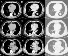
          The aim of this single-centre, observational, retrospective study is to find a correlation using Radiomics between the analysis of CT texture features of primary lesion of neuroendocrine (NET) lung cancer subtypes (typical and atypical carcinoids, large and small cell neuroendocrine carcinoma), Ki-67 index and the presence of lymph nodal mediastinal metastases.

          Methods

          Twenty-seven patients (11 males and 16 females, aged between 48 and 81 years old—average age of 70,4 years) with histological diagnosis of pulmonary NET with known Ki-67 status and metastases who have performed pre-treatment CT in our department were included. All examinations were performed with the same CT scan (Sensation 16-slice, Siemens). The study protocol was a baseline scan followed by 70 s delay acquisition after administration of intravenous contrast medium. After segmentation of primary lesions, quantitative texture parameters of first and higher orders were extracted. Statistics nonparametric tests and linear correlation tests were conducted to evaluate the relationship between different textural characteristics and tumour subtypes.

          Results

          Statistically significant ( p < 0.05) differences were seen in post-contrast enhanced CT in multiple first and higher-order extracted parameters regarding the correlation with classes of Ki-67 index values. Statistical analysis for direct acquisitions was not significant. Concerning the correlation with the presence of metastases, one histogram feature (Skewness) and one feature included in the Gray-Level Co-occurrence Matrix (ClusterShade) were significant on contrast-enhanced CT only.

          Conclusions

          CT texture analysis may be used as a valid tool for predicting the subtype of lung NET and its aggressiveness.

          Related collections

          Most cited references33

          • Record: found
          • Abstract: found
          • Article: not found

          Radiomics: Images Are More than Pictures, They Are Data

          This report describes the process of radiomics, its challenges, and its potential power to facilitate better clinical decision making, particularly in the care of patients with cancer.
            Bookmark
            • Record: found
            • Abstract: found
            • Article: not found

            Computational Radiomics System to Decode the Radiographic Phenotype

            Radiomics aims to quantify phenotypic characteristics on medical imaging through the use of automated algorithms. Radiomic artificial intelligence (AI) technology, either based on engineered hard-coded algorithms or deep learning methods, can be used to develop non-invasive imaging-based biomarkers. However, lack of standardized algorithm definitions and image processing severely hampers reproducibility and comparability of results. To address this issue, we developed PyRadiomics , a flexible open-source platform capable of extracting a large panel of engineered features from medical images. PyRadiomics is implemented in Python and can be used standalone or using 3D-Slicer. Here, we discuss the workflow and architecture of PyRadiomics and demonstrate its application in characterizing lung-lesions. Source code, documentation, and examples are publicly available at www.radiomics.io . With this platform, we aim to establish a reference standard for radiomic analyses, provide a tested and maintained resource, and to grow the community of radiomic developers addressing critical needs in cancer research.
              Bookmark
              • Record: found
              • Abstract: found
              • Article: not found

              3D Slicer as an image computing platform for the Quantitative Imaging Network.

              Quantitative analysis has tremendous but mostly unrealized potential in healthcare to support objective and accurate interpretation of the clinical imaging. In 2008, the National Cancer Institute began building the Quantitative Imaging Network (QIN) initiative with the goal of advancing quantitative imaging in the context of personalized therapy and evaluation of treatment response. Computerized analysis is an important component contributing to reproducibility and efficiency of the quantitative imaging techniques. The success of quantitative imaging is contingent on robust analysis methods and software tools to bring these methods from bench to bedside. 3D Slicer is a free open-source software application for medical image computing. As a clinical research tool, 3D Slicer is similar to a radiology workstation that supports versatile visualizations but also provides advanced functionality such as automated segmentation and registration for a variety of application domains. Unlike a typical radiology workstation, 3D Slicer is free and is not tied to specific hardware. As a programming platform, 3D Slicer facilitates translation and evaluation of the new quantitative methods by allowing the biomedical researcher to focus on the implementation of the algorithm and providing abstractions for the common tasks of data communication, visualization and user interface development. Compared to other tools that provide aspects of this functionality, 3D Slicer is fully open source and can be readily extended and redistributed. In addition, 3D Slicer is designed to facilitate the development of new functionality in the form of 3D Slicer extensions. In this paper, we present an overview of 3D Slicer as a platform for prototyping, development and evaluation of image analysis tools for clinical research applications. To illustrate the utility of the platform in the scope of QIN, we discuss several use cases of 3D Slicer by the existing QIN teams, and we elaborate on the future directions that can further facilitate development and validation of imaging biomarkers using 3D Slicer. Copyright © 2012 Elsevier Inc. All rights reserved.
                Bookmark

                Author and article information

                Contributors
                dilettacozzi@gmail.com
                eleonora.bicci92@gmail.com
                edoardocavigli@yahoo.it
                ginevra.danti@gmail.com
                silvia.bettarini@unifi.it
                paolo.tortoli@unifi.it
                mazzoniln@gmail.com
                busonis@aou-careggi.toscana.it
                pradella3@yahoo.it
                vmiele@sirm.org
                Journal
                Radiol Med
                Radiol Med
                La Radiologia Medica
                Springer Milan (Milan )
                0033-8362
                1826-6983
                10 May 2022
                10 May 2022
                2022
                : 127
                : 6
                : 609-615
                Affiliations
                [1 ]GRID grid.24704.35, ISNI 0000 0004 1759 9494, Department of Emergency Radiology, , Careggi University Hospital, ; Largo Brambilla 3, 50134 Florence, Italy
                [2 ]Italian Society of Medical and Interventional Radiology (SIRM), SIRM Foundation, 20122 Milan, Italy
                [3 ]GRID grid.24704.35, ISNI 0000 0004 1759 9494, Department of Health Physics, , Careggi University Hospital, ; Largo Brambilla 3, 50134 Florence, Italy
                [4 ]Department of Health Physics, AUSL Toscana Centro, Via Ciliegiole 97, 51100 Pistoia, Italy
                Author information
                http://orcid.org/0000-0001-7028-9047
                http://orcid.org/0000-0002-2078-0222
                http://orcid.org/0000-0001-6307-1621
                http://orcid.org/0000-0001-5310-1233
                http://orcid.org/0000-0002-3629-7278
                http://orcid.org/0000-0001-7453-007X
                http://orcid.org/0000-0002-7848-1567
                Article
                1494
                10.1007/s11547-022-01494-5
                9130162
                35538389
                a33a8bf0-c9d9-4460-bef7-862801140ed8
                © The Author(s) 2022

                Open AccessThis article is licensed under a Creative Commons Attribution 4.0 International License, which permits use, sharing, adaptation, distribution and reproduction in any medium or format, as long as you give appropriate credit to the original author(s) and the source, provide a link to the Creative Commons licence, and indicate if changes were made. The images or other third party material in this article are included in the article's Creative Commons licence, unless indicated otherwise in a credit line to the material. If material is not included in the article's Creative Commons licence and your intended use is not permitted by statutory regulation or exceeds the permitted use, you will need to obtain permission directly from the copyright holder. To view a copy of this licence, visit http://creativecommons.org/licenses/by/4.0/.

                History
                : 25 January 2022
                : 14 April 2022
                Categories
                Chest Radiology
                Custom metadata
                © Italian Society of Medical Radiology 2022

                lung carcinoids,computed tomography,radiomics,ki-67
                lung carcinoids, computed tomography, radiomics, ki-67

                Comments

                Comment on this article