17
views
0
recommends
+1 Recommend
0 collections
    0
    shares
      • Record: found
      • Abstract: found
      • Article: found
      Is Open Access

      Assessment of 25-Year Survival of Women With Estrogen Receptor–Positive/ ERBB2-Negative Breast Cancer Treated With and Without Tamoxifen Therapy : A Secondary Analysis of Data From the Stockholm Tamoxifen Randomized Clinical Trial

      research-article

      Read this article at

      Bookmark
          There is no author summary for this article yet. Authors can add summaries to their articles on ScienceOpen to make them more accessible to a non-specialist audience.

          Key Points

          Question

          Are clinically used markers of breast cancer, such as tumor size, tumor grade, progesterone receptor status, and Ki-67 status, independently associated with 25-year survival and tamoxifen treatment benefit among patients with breast cancer?

          Findings

          In this secondary analysis of data from 565 postmenopausal women with lymph node–negative, estrogen receptor–positive, and ERBB2-negative breast cancer who participated in the Stockholm tamoxifen randomized clinical trial (STO-3), tumor size and tumor grade were significantly associated with long-term (25-year) survival. A significant tamoxifen treatment benefit was observed among patients with larger tumors, lower tumor grades, and progesterone receptor–positive tumors.

          Meaning

          This study’s findings suggest that tumor size and tumor grade is associated with long-term survival, and patients with larger tumors, lower tumor grades, and progesterone receptor–positive status experienced significant treatment benefit with receipt of tamoxifen therapy.

          Abstract

          Importance

          Clinically used breast cancer markers, such as tumor size, tumor grade, progesterone receptor (PR) status, and Ki-67 status, are known to be associated with short-term survival, but the association of these markers with long-term (25-year) survival is unclear.

          Objective

          To assess the association of clinically used breast cancer markers with long-term survival and treatment benefit among postmenopausal women with lymph node–negative, estrogen receptor [ER]–positive and ERBB2-negative breast cancer who received tamoxifen therapy.

          Design, Setting, and Participants

          This study was a secondary analysis of data from a subset of 565 women with ER-positive/ ERBB2-negative breast cancer who participated in the Stockholm tamoxifen (STO-3) randomized clinical trial. The STO-3 clinical trial was conducted from 1976 to 1990 and comprised 1780 postmenopausal women with lymph node–negative breast cancer who were randomized to receive adjuvant tamoxifen therapy or no endocrine therapy. Complete 25-year follow-up data through December 31, 2016, were obtained from Swedish national registers. Immunohistochemical markers were reannotated in 2014. Data were analyzed from April to December 2020.

          Interventions

          Patients in the original STO-3 clinical trial were randomized to receive 2 years of tamoxifen therapy vs no endocrine therapy. In 1983, patients who received tamoxifen therapy without cancer recurrence during the 2-year treatment and who consented to continued participation in the STO-3 study were further randomized to receive 3 additional years of tamoxifen therapy or no endocrine therapy.

          Main Outcomes and Measures

          Distant recurrence–free interval (DRFI) by clinically used breast cancer markers was assessed using Kaplan-Meier and multivariable Cox proportional hazards analyses adjusted for age, period of primary diagnosis, tumor size (T1a and T1b [T1a/b], T1c, and T2), tumor grade (1-3), PR status (positive vs negative), Ki-67 status (low vs medium to high), and STO-3 clinical trial arm (tamoxifen treatment vs no adjuvant treatment). A recursive partitioning analysis was performed to evaluate which markers were able to best estimate long-term DRFI.

          Results

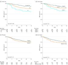
          The study population comprised 565 postmenopausal women (mean [SD] age, 62.0 [5.3] years) with lymph node–negative, ER-positive/ ERBB2-negative breast cancer. A statistically significant difference in long-term DRFI was observed by tumor size (88% for T1a/b vs 76% for T1c vs 63% for T2 tumors; log-rank P < .001) and tumor grade (81% for grade 1 vs 77% for grade 2 vs 65% for grade 3 tumors; log-rank P = .02) but not by PR status or Ki-67 status. Patients with smaller tumors (hazard ratio [HR], 0.31 [95% CI, 0.17-0.55] for T1a/b tumors and 0.58 [95% CI, 0.38-0.88] for T1c tumors) and grade 1 tumors (HR, 0.48; 95% CI, 0.24-0.95) experienced a significant reduction in the long-term risk of distant recurrence compared with patients with larger (T2) tumors and grade 3 tumors, respectively. A significant tamoxifen treatment benefit was observed among patients with larger tumors (HR, 0.53 [95% CI, 0.32-0.89] for T1c tumors and 0.34 [95% CI, 0.16-0.73] for T2 tumors), lower tumor grades (HR, 0.24 [95% CI, 0.07-0.82] for grade 1 tumors and 0.50 [95% CI, 0.31-0.80] for grade 2 tumors), and PR-positive status (HR, 0.38; 95% CI, 0.24-0.62). The recursive partitioning analysis revealed that tumor size was the most important characteristic associated with long-term survival, followed by clinical trial arm among patients with larger tumors.

          Conclusions and Relevance

          This secondary analysis of data from the STO-3 clinical trial indicated that, among the selected subgroup of patients, tumor size followed by tumor grade were the markers most significantly associated with long-term survival. Furthermore, a significant long-term tamoxifen treatment benefit was observed among patients with larger tumors, lower tumor grades, and PR-positive tumors.

          Abstract

          This secondary analysis of the Stockholm tamoxifen clinical trial examines the association of clinical breast cancer markers with 25-year survival and tamoxifen treatment benefit among postmenopausal women with lymph node–negative ER-positive/ ERBB2-negative breast cancer.

          Related collections

          Most cited references29

          • Record: found
          • Abstract: found
          • Article: not found

          20-Year Risks of Breast-Cancer Recurrence after Stopping Endocrine Therapy at 5 Years.

          The administration of endocrine therapy for 5 years substantially reduces recurrence rates during and after treatment in women with early-stage, estrogen-receptor (ER)-positive breast cancer. Extending such therapy beyond 5 years offers further protection but has additional side effects. Obtaining data on the absolute risk of subsequent distant recurrence if therapy stops at 5 years could help determine whether to extend treatment.
            Bookmark
            • Record: found
            • Abstract: found
            • Article: found

            Relevance of breast cancer hormone receptors and other factors to the efficacy of adjuvant tamoxifen: patient-level meta-analysis of randomised trials

            Summary Background As trials of 5 years of tamoxifen in early breast cancer mature, the relevance of hormone receptor measurements (and other patient characteristics) to long-term outcome can be assessed increasingly reliably. We report updated meta-analyses of the trials of 5 years of adjuvant tamoxifen. Methods We undertook a collaborative meta-analysis of individual patient data from 20 trials (n=21 457) in early breast cancer of about 5 years of tamoxifen versus no adjuvant tamoxifen, with about 80% compliance. Recurrence and death rate ratios (RRs) were from log-rank analyses by allocated treatment. Findings In oestrogen receptor (ER)-positive disease (n=10 645), allocation to about 5 years of tamoxifen substantially reduced recurrence rates throughout the first 10 years (RR 0·53 [SE 0·03] during years 0–4 and RR 0·68 [0·06] during years 5–9 [both 2p<0·00001]; but RR 0·97 [0·10] during years 10–14, suggesting no further gain or loss after year 10). Even in marginally ER-positive disease (10–19 fmol/mg cytosol protein) the recurrence reduction was substantial (RR 0·67 [0·08]). In ER-positive disease, the RR was approximately independent of progesterone receptor status (or level), age, nodal status, or use of chemotherapy. Breast cancer mortality was reduced by about a third throughout the first 15 years (RR 0·71 [0·05] during years 0–4, 0·66 [0·05] during years 5–9, and 0·68 [0·08] during years 10–14; p<0·0001 for extra mortality reduction during each separate time period). Overall non-breast-cancer mortality was little affected, despite small absolute increases in thromboembolic and uterine cancer mortality (both only in women older than 55 years), so all-cause mortality was substantially reduced. In ER-negative disease, tamoxifen had little or no effect on breast cancer recurrence or mortality. Interpretation 5 years of adjuvant tamoxifen safely reduces 15-year risks of breast cancer recurrence and death. ER status was the only recorded factor importantly predictive of the proportional reductions. Hence, the absolute risk reductions produced by tamoxifen depend on the absolute breast cancer risks (after any chemotherapy) without tamoxifen. Funding Cancer Research UK, British Heart Foundation, and Medical Research Council.
              Bookmark
              • Record: found
              • Abstract: found
              • Article: not found

              Relation of tumor size, lymph node status, and survival in 24,740 breast cancer cases.

              Two of the most important prognostic indicators for breast cancer are tumor size and extent of axillary lymph node involvement. Data on 24,740 cases recorded in the Surveillance, Epidemiology, and End Results (SEER) Program of the National Cancer Institute were used to evaluate the breast cancer survival experience in a representative sample of women from the United States. Actuarial (life table) methods were used to investigate the 5-year relative survival rates in cases with known operative/pathologic axillary lymph node status and primary tumor diameter. Survival rates varied from 45.5% for tumor diameters equal to or greater than 5 cm with positive axillary nodes to 96.3% for tumors less than 2 cm and with no involved nodes. The relation between tumor size and lymph node status was investigated in detail. Tumor diameter and lymph node status were found to act as independent but additive prognostic indicators. As tumor size increased, survival decreased regardless of lymph node status; and as lymph node involvement increased, survival status also decreased regardless of tumor size. A linear relation was found between tumor diameter and the percent of cases with positive lymph node involvement. The results of our analyses suggest that disease progression to distant sites does not occur exclusively via the axillary lymph nodes, but rather that lymph node status serves as an indicator of the tumor's ability to spread.
                Bookmark

                Author and article information

                Journal
                JAMA Netw Open
                JAMA Netw Open
                JAMA Netw Open
                JAMA Network Open
                American Medical Association
                2574-3805
                30 June 2021
                June 2021
                30 June 2021
                : 4
                : 6
                : e2114904
                Affiliations
                [1 ]Department of Oncology and Pathology, Karolinska Institutet and University Hospital, Stockholm, Sweden
                [2 ]Department of Oncology, Institute of Clinical Sciences, Sahlgrenska Academy, Gothenburg, Sweden
                [3 ]Department of Medicine, Southern Älvsborg Hospital, Borås, Sweden
                [4 ]Department of Biosciences and Nutrition, Karolinska Institutet, Stockholm, Sweden
                [5 ]Department of Surgery, University of California, San Francisco, San Francisco
                [6 ]Department of Biomedical and Clinical Sciences, Linköping University, Linköping, Sweden
                [7 ]Department of Oncology, Linköping University, Linköping, Sweden
                [8 ]Department of Medicine, University of California, San Francisco, San Francisco
                Author notes
                Article Information
                Accepted for Publication: April 6, 2021.
                Published: June 30, 2021. doi:10.1001/jamanetworkopen.2021.14904
                Open Access: This is an open access article distributed under the terms of the CC-BY License. © 2021 Dar H et al. JAMA Network Open.
                Corresponding Authors: Linda S. Lindström, MSc, PhD ( linda.lindstrom@ 123456ki.se ), and Huma Dar, MSc ( huma.dar@ 123456ki.se ), Department of Oncology and Pathology, Karolinska Institutet and University Hospital, Visionsgatan 4 Bioclinicum, Visionsgatan 4, 171 64 Stockholm, Sweden.
                Author Contributions: Dr Lindström had full access to all of the data in the study and takes responsibility for the integrity of the data and the accuracy of the data analysis.
                Concept and design: Dar, B. Nordenskjöld, Stål, Esserman, Fornander, Lindström.
                Acquisition, analysis, or interpretation of data: All authors.
                Drafting of the manuscript: Dar, A. Nordenskjöld, B. Nordenskjöld, Esserman, Lindström.
                Critical revision of the manuscript for important intellectual content: All authors.
                Statistical analysis: Dar, Johansson, Iftimi, Lindström.
                Obtained funding: B. Nordenskjöld, Lindström.
                Administrative, technical, or material support: B. Nordenskjöld, Esserman, Fornander, Lindström.
                Supervision: Johansson, Stål, Fornander, Lindström.
                Conflict of Interest Disclosures: Dr Yau reported receiving grants from the National Cancer Institute of the National Institutes of Health during the conduct of the study and personal fees from NantOmics outside the submitted work. Dr Esserman reported receiving grants from Merck & Co and Quantum Leap Healthcare Collaborative and personal fees from the Blue Cross Blue Shield Medical Advisory Panel outside the submitted work. Dr Fornander reported receiving grants from the Stockholm Cancer Society during the conduct of the study. No other disclosures were reported.
                Funding/Support: This work was supported by grant 2020-02466 (Dr Lindström) from the Swedish Research Council (Vetenskapsrådet); grant 2019-00477 from the Swedish Research Council for Health, Working life and Welfare (FORTE); grants 180385 (Dr Stål) and 190140Pj01H (Dr Lindström) from the Swedish Cancer Society (Cancerfonden); and funding from Gösta Milton Donation Fund (Dr Lindström).
                Role of the Funder/Sponsor: The funders had no role in the design and conduct of the study; collection, management, analysis, and interpretation of the data; preparation, review, or approval of the manuscript; and decision to submit the manuscript for publication.
                Data Sharing Statement: See Supplement 2.
                Additional Contributions: The authors thank all of the patients in this study, their treating physicians, and the collaborating hospitals, in addition to the pathology departments.
                Article
                zoi210451
                10.1001/jamanetworkopen.2021.14904
                8246315
                34190995
                a20425ef-6491-4f82-b68d-ef4fe805b848
                Copyright 2021 Dar H et al. JAMA Network Open.

                This is an open access article distributed under the terms of the CC-BY License.

                History
                : 12 January 2021
                : 6 April 2021
                Categories
                Research
                Original Investigation
                Featured
                Online Only
                Oncology

                Comments

                Comment on this article