4
views
0
recommends
+1 Recommend
0 collections
    0
    shares
      • Record: found
      • Abstract: found
      • Article: found
      Is Open Access

      Improving CBCT image quality to the CT level using RegGAN in esophageal cancer adaptive radiotherapy

      research-article

      Read this article at

      Bookmark
          There is no author summary for this article yet. Authors can add summaries to their articles on ScienceOpen to make them more accessible to a non-specialist audience.

          Abstract

          Objective

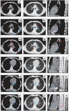
          This study aimed to improve the image quality and CT Hounsfield unit accuracy of daily cone-beam computed tomography (CBCT) using registration generative adversarial networks (RegGAN) and apply synthetic CT (sCT) images to dose calculations in radiotherapy.

          Methods

          The CBCT/planning CT images of 150 esophageal cancer patients undergoing radiotherapy were used for training (120 patients) and testing (30 patients). An unsupervised deep-learning method, the 2.5D RegGAN model with an adaptively trained registration network, was proposed, through which sCT images were generated. The quality of deep-learning-generated sCT images was quantitatively compared to the reference deformed CT (dCT) image using mean absolute error (MAE), root mean square error (RMSE) of Hounsfield units (HU), and peak signal-to-noise ratio (PSNR). The dose calculation accuracy was further evaluated for esophageal cancer radiotherapy plans, and the same plans were calculated on dCT, CBCT, and sCT images.

          Results

          The quality of sCT images produced by RegGAN was significantly improved compared to the original CBCT images. ReGAN achieved image quality in the testing patients with MAE sCT vs. CBCT: 43.7 ± 4.8 vs. 80.1 ± 9.1; RMSE sCT vs. CBCT: 67.2 ± 12.4 vs. 124.2 ± 21.8; and PSNR sCT vs. CBCT: 27.9 ± 5.6 vs. 21.3 ± 4.2. The sCT images generated by the RegGAN model showed superior accuracy on dose calculation, with higher gamma passing rates (93.3 ± 4.4, 90.4 ± 5.2, and 84.3 ± 6.6) compared to original CBCT images (89.6 ± 5.7, 85.7 ± 6.9, and 72.5 ± 12.5) under the criteria of 3 mm/3%, 2 mm/2%, and 1 mm/1%, respectively.

          Conclusion

          The proposed deep-learning RegGAN model seems promising for generation of high-quality sCT images from stand-alone thoracic CBCT images in an efficient way and thus has the potential to support CBCT-based esophageal cancer adaptive radiotherapy.

          Related collections

          Most cited references27

          • Record: found
          • Abstract: not found
          • Conference Proceedings: not found

          Image-to-Image Translation with Conditional Adversarial Networks

            Bookmark
            • Record: found
            • Abstract: not found
            • Conference Proceedings: not found

            Unpaired Image-to-Image Translation Using Cycle-Consistent Adversarial Networks

              Bookmark
              • Record: found
              • Abstract: found
              • Article: not found

              Improved scatter correction using adaptive scatter kernel superposition.

              Accurate scatter correction is required to produce high-quality reconstructions of x-ray cone-beam computed tomography (CBCT) scans. This paper describes new scatter kernel superposition (SKS) algorithms for deconvolving scatter from projection data. The algorithms are designed to improve upon the conventional approach whose accuracy is limited by the use of symmetric kernels that characterize the scatter properties of uniform slabs. To model scatter transport in more realistic objects, nonstationary kernels, whose shapes adapt to local thickness variations in the projection data, are proposed. Two methods are introduced: (1) adaptive scatter kernel superposition (ASKS) requiring spatial domain convolutions and (2) fast adaptive scatter kernel superposition (fASKS) where, through a linearity approximation, convolution is efficiently performed in Fourier space. The conventional SKS algorithm, ASKS, and fASKS, were tested with Monte Carlo simulations and with phantom data acquired on a table-top CBCT system matching the Varian On-Board Imager (OBI). All three models accounted for scatter point-spread broadening due to object thickening, object edge effects, detector scatter properties and an anti-scatter grid. Hounsfield unit (HU) errors in reconstructions of a large pelvis phantom with a measured maximum scatter-to-primary ratio over 200% were reduced from -90 ± 58 HU (mean ± standard deviation) with no scatter correction to 53 ± 82 HU with SKS, to 19 ± 25 HU with fASKS and to 13 ± 21 HU with ASKS. HU accuracies and measured contrast were similarly improved in reconstructions of a body-sized elliptical Catphan phantom. The results show that the adaptive SKS methods offer significant advantages over the conventional scatter deconvolution technique.
                Bookmark

                Author and article information

                Contributors
                zhou.yongkang@zs-hospital.sh.cn
                Journal
                Strahlenther Onkol
                Strahlenther Onkol
                Strahlentherapie Und Onkologie
                Springer Berlin Heidelberg (Berlin/Heidelberg )
                0179-7158
                1439-099X
                23 January 2023
                23 January 2023
                2023
                : 199
                : 5
                : 485-497
                Affiliations
                [1 ]GRID grid.16821.3c, ISNI 0000 0004 0368 8293, Department of Radiation Oncology, Shanghai Chest Hospital, , Shanghai Jiaotong University, ; Shanghai, China
                [2 ]GRID grid.410587.f, Department of Radiotherapy, Shandong Cancer Hospital and Institute, , Shandong First Medical University and Shandong Academy of Medical Sciences, ; Jinan, China
                [3 ]Manteia Tech, Xiamen, China
                [4 ]GRID grid.8547.e, ISNI 0000 0001 0125 2443, Institute of Modern Physics, , Fudan University, ; Shanghai, China
                [5 ]GRID grid.8547.e, ISNI 0000 0001 0125 2443, Department of Radiation Oncology, Zhongshan Hospital, , Fudan University, ; Shanghai, China
                Author information
                http://orcid.org/0000-0001-5722-0432
                Article
                2039
                10.1007/s00066-022-02039-5
                10133081
                36688953
                9ff43c0c-ac71-47e5-ac54-454bc10370ca
                © The Author(s) 2023

                Open Access This article is licensed under a Creative Commons Attribution 4.0 International License, which permits use, sharing, adaptation, distribution and reproduction in any medium or format, as long as you give appropriate credit to the original author(s) and the source, provide a link to the Creative Commons licence, and indicate if changes were made. The images or other third party material in this article are included in the article’s Creative Commons licence, unless indicated otherwise in a credit line to the material. If material is not included in the article’s Creative Commons licence and your intended use is not permitted by statutory regulation or exceeds the permitted use, you will need to obtain permission directly from the copyright holder. To view a copy of this licence, visit http://creativecommons.org/licenses/by/4.0/.

                History
                : 5 September 2022
                : 4 December 2022
                Categories
                Original Article
                Custom metadata
                © Springer-Verlag GmbH Germany, part of Springer Nature 2023

                Oncology & Radiotherapy
                deep learning,diagnostic imaging,neural networks, computer,organs at risk,artifacts

                Comments

                Comment on this article