37
views
0
recommends
+1 Recommend
0 collections
    0
    shares
      • Record: found
      • Abstract: found
      • Article: found
      Is Open Access

      Atlastins remodel the endoplasmic reticulum for selective autophagy

      research-article

      Read this article at

      Bookmark
          There is no author summary for this article yet. Authors can add summaries to their articles on ScienceOpen to make them more accessible to a non-specialist audience.

          Abstract

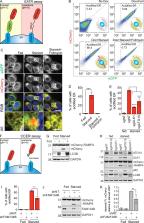
          Multiple ER-phagy receptors have been reported recently, but other mediators or regulators have remained elusive. Liang et al. developed two ER-phagy–specific reporter assays and showed that Atlastins, a family of ER surface GTPases, are positive regulators of ER-phagy that act downstream of an ER-phagy receptor, FAM134B.

          Abstract

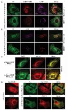
          Specific receptors are required for the autophagic degradation of endoplasmic reticulum (ER), known as ER-phagy. However, little is known about how the ER is remodeled and separated for packaging into autophagosomes. We developed two ER-phagy–specific reporter systems and found that Atlastins are key positive effectors and also targets of ER-phagy. Atlastins are ER-resident GTPases involved in ER membrane morphology, and Atlastin-depleted cells have decreased ER-phagy under starvation conditions. Atlastin’s role in ER-phagy requires a functional GTPase domain and proper ER localization, both of which are also involved in ER architecture. The three Atlastin family members functionally compensate for one another during ER-phagy and may form heteromeric complexes with one another. We further find that Atlastins act downstream of the FAM134B ER-phagy receptor, such that depletion of Atlastins represses ER-autophagy induced by the overexpression of FAM134B. We propose that during ER-phagy, Atlastins remodel ER membrane to separate pieces of FAM134B-marked ER for efficient autophagosomal engulfment.

          Related collections

          Most cited references20

          • Record: found
          • Abstract: found
          • Article: not found

          A class of membrane proteins shaping the tubular endoplasmic reticulum.

          How is the characteristic shape of a membrane bound organelle achieved? We have used an in vitro system to address the mechanism by which the tubular network of the endoplasmic reticulum (ER) is generated and maintained. Based on the inhibitory effect of sulfhydryl reagents and antibodies, network formation in vitro requires the integral membrane protein Rtn4a/NogoA, a member of the ubiquitous reticulon family. Both in yeast and mammalian cells, the reticulons are largely restricted to the tubular ER and are excluded from the continuous sheets of the nuclear envelope and peripheral ER. Upon overexpression, the reticulons form tubular membrane structures. The reticulons interact with DP1/Yop1p, a conserved integral membrane protein that also localizes to the tubular ER. These proteins share an unusual hairpin topology in the membrane. The simultaneous absence of the reticulons and Yop1p in S. cerevisiae results in disrupted tubular ER. We propose that these "morphogenic" proteins partition into and stabilize highly curved ER membrane tubules.
            Bookmark
            • Record: found
            • Abstract: found
            • Article: found
            Is Open Access

            Full length RTN3 regulates turnover of tubular endoplasmic reticulum via selective autophagy

            The turnover of endoplasmic reticulum (ER) ensures the correct biological activity of its distinct domains. In mammalian cells, the ER is degraded via a selective autophagy pathway (ER-phagy), mediated by two specific receptors: FAM134B, responsible for the turnover of ER sheets and SEC62 that regulates ER recovery following stress. Here, we identified reticulon 3 (RTN3) as a specific receptor for the degradation of ER tubules. Oligomerization of the long isoform of RTN3 is sufficient to trigger fragmentation of ER tubules. The long N-terminal region of RTN3 contains several newly identified LC3-interacting regions (LIR). Binding to LC3s/GABARAPs is essential for the fragmentation of ER tubules and their delivery to lysosomes. RTN3-mediated ER-phagy requires conventional autophagy components, but is independent of FAM134B. None of the other reticulon family members have the ability to induce fragmentation of ER tubules during starvation. Therefore, we assign a unique function to RTN3 during autophagy. DOI: http://dx.doi.org/10.7554/eLife.25555.001
              Bookmark
              • Record: found
              • Abstract: found
              • Article: not found

              Cleaning up: ER-associated degradation to the rescue.

              All cellular proteins are subject to quality control "decisions," which help to prevent or delay a myriad of diseases. Quality control within the secretory pathway creates a special challenge, as aberrant polypeptides are recognized and returned to the cytoplasm for proteasomal degradation. This process is termed endoplasmic-reticulum (ER)-associated degradation (ERAD). Copyright © 2012 Elsevier Inc. All rights reserved.
                Bookmark

                Author and article information

                Journal
                J Cell Biol
                J. Cell Biol
                jcb
                jcb
                The Journal of Cell Biology
                Rockefeller University Press
                0021-9525
                1540-8140
                01 October 2018
                : 217
                : 10
                : 3354-3367
                Affiliations
                [1 ]Innovative Genomics Institute, University of California, Berkeley, Berkeley, CA
                [2 ]Department of Molecular and Cell Biology, University of California, Berkeley, Berkeley, CA
                Author notes
                Correspondence to Jacob E. Corn: jcorn@ 123456berkeley.edu
                Author information
                http://orcid.org/0000-0002-8774-6061
                http://orcid.org/0000-0002-7798-5309
                Article
                201804185
                10.1083/jcb.201804185
                6168278
                30143524
                9f22a81b-c1fe-4b81-874b-d2fea0011814
                © 2018 Liang et al.

                This article is distributed under the terms of an Attribution–Noncommercial–Share Alike–No Mirror Sites license for the first six months after the publication date (see http://www.rupress.org/terms/). After six months it is available under a Creative Commons License (Attribution–Noncommercial–Share Alike 4.0 International license, as described at https://creativecommons.org/licenses/by-nc-sa/4.0/).

                History
                : 26 April 2018
                : 15 June 2018
                : 11 July 2018
                Funding
                Funded by: National Institutes of Health, DOI https://doi.org/10.13039/100000002;
                Award ID: DP2-HL-141006
                Funded by: Gordon and Betty Moore Foundation, DOI https://doi.org/10.13039/100000936;
                Categories
                Research Articles
                Report
                35
                1
                22

                Cell biology
                Cell biology

                Comments

                Comment on this article

                scite_
                0
                0
                0
                0
                Smart Citations
                0
                0
                0
                0
                Citing PublicationsSupportingMentioningContrasting
                View Citations

                See how this article has been cited at scite.ai

                scite shows how a scientific paper has been cited by providing the context of the citation, a classification describing whether it supports, mentions, or contrasts the cited claim, and a label indicating in which section the citation was made.

                Similar content27

                Cited by70

                Most referenced authors604