0
views
0
recommends
+1 Recommend
0 collections
    0
    shares
      • Record: found
      • Abstract: found
      • Article: found
      Is Open Access

      Efficacy of Coronary Calcium Score in Predicting Coronary Artery Morphology in Patients With Obstructive Coronary Artery Disease

      research-article
      , MD a , b , , , MBChB a , , , MBChB, PhD a , c , , PhD a , d , , MBChB a , c , , MBChB a , , MD e , , MD, PhD f , , MSc g , h , , PhD h , , PhD g , h , , PhD h , , MD, PhD i , j , , MD a , k , , MD, PhD a , c , , MSc, PhD l , , MD, PhD a , c , , MD, PhD a , c ,
      Journal of the Society for Cardiovascular Angiography & Interventions
      Elsevier
      coronary artery disease, coronary calcium score, coronary computed tomography angiography, near-infrared spectroscopy-intravascular ultrasound, risk stratification, vulnerable plaque

      Read this article at

      Bookmark
          There is no author summary for this article yet. Authors can add summaries to their articles on ScienceOpen to make them more accessible to a non-specialist audience.

          Abstract

          Background

          Coronary artery calcium score (CACS) is an established marker of coronary artery disease (CAD) and has been extensively used to stratify risk in asymptomatic individuals. However, the value of CACS in predicting plaque morphology in patients with advanced CAD is less established. The present analysis aims to assess the association between CACS and plaque characteristics detected by near-infrared spectroscopy-intravascular ultrasound (NIRS-IVUS) imaging in patients with obstructive CAD.

          Methods

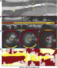
          Seventy patients with obstructive CAD underwent coronary computed tomography angiography (CTA) and 3-vessel NIRS-IVUS imaging were included in the present analysis. The CTA data were used to measure the CACS in the entire coronary tree and the segments assessed by NIRS-IVUS, and these estimations were associated with the NIRS-IVUS measurements at a patient and segment level.

          Results

          In total, 65 patients (188 segments) completed the study protocol and were included in the analysis. A weak correlation was noted between the CACS, percent atheroma volume (r = 0.271, P = .002), and the calcific burden measured by NIRS-IVUS (r = 0.648, P < .001) at patient-level analysis. Conversely, there was no association between the CACS and the lipid content, or the incidence of high-risk plaques detected by NIRS. Linear regression analysis at the segment level demonstrated an association between the CACS and the total atheroma volume (coefficient, 0.087; 95% CI, 0.024-0.149; P = .008) and the calcific burden (coefficient, 0.117; 95% CI, 0.048-0.186; P = .001), but there was no association between the lipid content or the incidence of high-risk lesions.

          Conclusions

          In patients with obstructive CAD, the CACS is not associated with the lipid content or plaque phenotypes. These findings indicate that the CACS may have a limited value for screening or stratifying cardiovascular risk in symptomatic patients with a high probability of CAD.

          Related collections

          Most cited references35

          • Record: found
          • Abstract: not found
          • Article: not found

          2021 ESC Guidelines on cardiovascular disease prevention in clinical practice

            Bookmark
            • Record: found
            • Abstract: found
            • Article: not found

            A prospective natural-history study of coronary atherosclerosis.

            Atherosclerotic plaques that lead to acute coronary syndromes often occur at sites of angiographically mild coronary-artery stenosis. Lesion-related risk factors for such events are poorly understood. In a prospective study, 697 patients with acute coronary syndromes underwent three-vessel coronary angiography and gray-scale and radiofrequency intravascular ultrasonographic imaging after percutaneous coronary intervention. Subsequent major adverse cardiovascular events (death from cardiac causes, cardiac arrest, myocardial infarction, or rehospitalization due to unstable or progressive angina) were adjudicated to be related to either originally treated (culprit) lesions or untreated (nonculprit) lesions. The median follow-up period was 3.4 years. The 3-year cumulative rate of major adverse cardiovascular events was 20.4%. Events were adjudicated to be related to culprit lesions in 12.9% of patients and to nonculprit lesions in 11.6%. Most nonculprit lesions responsible for follow-up events were angiographically mild at baseline (mean [±SD] diameter stenosis, 32.3±20.6%). However, on multivariate analysis, nonculprit lesions associated with recurrent events were more likely than those not associated with recurrent events to be characterized by a plaque burden of 70% or greater (hazard ratio, 5.03; 95% confidence interval [CI], 2.51 to 10.11; P<0.001) or a minimal luminal area of 4.0 mm(2) or less (hazard ratio, 3.21; 95% CI, 1.61 to 6.42; P=0.001) or to be classified on the basis of radiofrequency intravascular ultrasonography as thin-cap fibroatheromas (hazard ratio, 3.35; 95% CI, 1.77 to 6.36; P<0.001). In patients who presented with an acute coronary syndrome and underwent percutaneous coronary intervention, major adverse cardiovascular events occurring during follow-up were equally attributable to recurrence at the site of culprit lesions and to nonculprit lesions. Although nonculprit lesions that were responsible for unanticipated events were frequently angiographically mild, most were thin-cap fibroatheromas or were characterized by a large plaque burden, a small luminal area, or some combination of these characteristics, as determined by gray-scale and radiofrequency intravascular ultrasonography. (Funded by Abbott Vascular and Volcano; ClinicalTrials.gov number, NCT00180466.).
              Bookmark
              • Record: found
              • Abstract: found
              • Article: not found

              Long-term prognosis associated with coronary calcification: observations from a registry of 25,253 patients.

              The purpose of this study was to develop risk-adjusted multivariable models that include risk factors and coronary artery calcium (CAC) scores measured with electron-beam tomography in asymptomatic patients for the prediction of all-cause mortality. Several smaller studies have documented the efficacy of CAC testing for assessment of cardiovascular risk. Larger studies with longer follow-up will lend strength to the hypothesis that CAC testing will improve outcomes, cost-effectiveness, and safety of primary prevention efforts. We used an observational outcome study of a cohort of 25,253 consecutive, asymptomatic individuals referred by their primary physician for CAC scanning to assess cardiovascular risk. Multivariable Cox proportional hazards models were developed to predict all-cause mortality. Risk-adjusted models incorporated traditional risk factors for coronary disease and CAC scores. The frequency of CAC scores was 44%, 14%, 20%, 13%, 6%, and 4% for scores of 0, 1 to 10, 11 to 100, 101 to 400, 401 to 1,000, and >1,000, respectively. During a mean follow-up of 6.8 +/- 3 years, the death rate was 2% (510 deaths). The CAC was an independent predictor of mortality in a multivariable model controlling for age, gender, ethnicity, and cardiac risk factors (model chi-square = 2,017, p 1,000, respectively (p 1,000 (p < 0.0001). This large observational data series shows that CAC provides independent incremental information in addition to traditional risk factors in the prediction of all-cause mortality.
                Bookmark

                Author and article information

                Contributors
                Journal
                J Soc Cardiovasc Angiogr Interv
                J Soc Cardiovasc Angiogr Interv
                Journal of the Society for Cardiovascular Angiography & Interventions
                Elsevier
                2772-9303
                26 March 2024
                March 2024
                26 March 2024
                : 3
                : 3Part B
                : 101308
                Affiliations
                [a ]Department of Cardiology, Barts Heart Centre, Barts Health NHS Trust, London, United Kingdom
                [b ]Division of Cardiology, Department of Internal Medicine, Tongji Hospital, Tongji Medical College, Huazhong University of Science and Technology, Wuhan, China
                [c ]Centre for Cardiovascular Medicine and Devices, William Harvey Research Institute, Queen Mary University of London, London, United Kingdom
                [d ]Institute of Health Informatics, University College London, London, United Kingdom
                [e ]Department of Cardiology, Faculty of Medicine, Yuzuncu Yil University Van, Van, Turkey
                [f ]Department of Cardiology, Xuzhou Third People’s Hospital, Xuzhou, China
                [g ]Division of Image Processing, Department of Radiology, Leiden University Medical Center, Leiden, the Netherlands
                [h ]Medis Medical Imaging Systems, Leiden, the Netherlands
                [i ]Faculty of Medicine, National Heart & Lung Institute, Imperial College London, London, United Kingdom
                [j ]Department of Cardiology, University of Galway, Galway, Ireland
                [k ]Institute of Cardiovascular Sciences, University College London, London, United Kingdom
                [l ]Department of Mechanical Engineering, University College London, London, United Kingdom
                Author notes
                []Corresponding author c.bourantas@ 123456gmail.com
                [†]

                Co-first authors.

                Article
                S2772-9303(24)00015-2 101308
                10.1016/j.jscai.2024.101308
                11307846
                9ef235d1-0022-406c-91e5-7271d3c2e2a4
                © 2024 The Author(s)

                This is an open access article under the CC BY-NC-ND license (http://creativecommons.org/licenses/by-nc-nd/4.0/).

                History
                : 2 January 2024
                : 11 January 2024
                : 12 January 2024
                Categories
                Original Research

                coronary artery disease,coronary calcium score,coronary computed tomography angiography,near-infrared spectroscopy-intravascular ultrasound,risk stratification,vulnerable plaque

                Comments

                Comment on this article