3
views
0
recommends
+1 Recommend
0 collections
    0
    shares
      • Record: found
      • Abstract: found
      • Article: found
      Is Open Access

      Differences in in vitro microglial accumulation of the energy metabolism tracers [ 18F]FDG and [ 18F]BCPP-EF during LPS- and IL4 stimulation

      research-article

      Read this article at

      Bookmark
          There is no author summary for this article yet. Authors can add summaries to their articles on ScienceOpen to make them more accessible to a non-specialist audience.

          Abstract

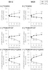
          The positron emission tomography probes 2-deoxy-2-[ 18F]fluoro-D-glucose ([ 18F]FDG) and 2- tert-butyl-4-chloro-5-{6-[2-(2-[ 18F]fluoroethoxy)-ethoxy]-pyridin-3-ylmethoxy}-2H-pyridazin-3-one ([ 18F]BCPP-EF) are designed to evaluate glycolysis and oxidative phosphorylation, respectively, and are both used to estimate neuronal activity. However, previous studies have shown a discrepancy in these probes’ accumulation in the compromised region, possibly due to the presence of activated microglia acting like deleterious or neuroprotective phenotypes. Hence, we evaluated lipopolysaccharide (LPS)- and interleukin 4 (IL4)-stimulated microglial uptake of [ 14C]2DG and [ 18F]BCPP-EF to give a new insight into the hypothesis that different uptake of [ 18F]FDG and [ 18F]BCPP-EF can be ascribed to the different metabolic pathways activated during microglial activation. LPS or IL4 stimulation increased the proinflammatory or anti-inflammatory marker gene expression in microglial cells. In LPS-stimulated cells, [ 14C]2DG uptake and glycolysis related gene expression were elevated, and [ 18F]BCPP-EF uptake was reduced. In IL4-stimulated cells, [ 18F]BCPP-EF uptake was increased, and [ 14C]2DG uptake was decreased. The expression of genes involved in glycolysis and mitochondrial complex I subunits was not changed by IL4 stimulation. The uptake of [ 14C]2DG and [ 18F]BCPP-EF differs in LPS- and IL4-stimulated polarized microglial cells. The present results suggest that the in vivo accumulation of metabolic tracers [ 18F]FDG and [ 18F]BCPP-EF can be influenced by the different aspects of neuroinflammation.

          Related collections

          Most cited references42

          • Record: found
          • Abstract: found
          • Article: found

          Alzheimer's disease.

          Although the prevalence of dementia continues to increase worldwide, incidence in the western world might have decreased as a result of better vascular care and improved brain health. Alzheimer's disease, the most prevalent cause of dementia, is still defined by the combined presence of amyloid and tau, but researchers are gradually moving away from the simple assumption of linear causality as proposed in the original amyloid hypothesis. Age-related, protective, and disease-promoting factors probably interact with the core mechanisms of the disease. Amyloid β42, and tau proteins are established core cerebrospinal biomarkers; novel candidate biomarkers include amyloid β oligomers and synaptic markers. MRI and fluorodeoxyglucose PET are established imaging techniques for diagnosis of Alzheimer's disease. Amyloid PET is gaining traction in the clinical arena, but validity and cost-effectiveness remain to be established. Tau PET might offer new insights and be of great help in differential diagnosis and selection of patients for trials. In the search for understanding the disease mechanism and keys to treatment, research is moving increasingly into the earliest phase of disease. Preclinical Alzheimer's disease is defined as biomarker evidence of Alzheimer's pathological changes in cognitively healthy individuals. Patients with subjective cognitive decline have been identified as a useful population in whom to look for preclinical Alzheimer's disease. Moderately positive results for interventions targeting several lifestyle factors in non-demented elderly patients and moderately positive interim results for lowering amyloid in pre-dementia Alzheimer's disease suggest that, ultimately, there will be a future in which specific anti-Alzheimer's therapy will be combined with lifestyle interventions targeting general brain health to jointly combat the disease. In this Seminar, we discuss the main developments in Alzheimer's research.
            Bookmark
            • Record: found
            • Abstract: found
            • Article: not found

            Hypothetical model of dynamic biomarkers of the Alzheimer's pathological cascade.

            Currently available evidence strongly supports the position that the initiating event in Alzheimer's disease (AD) is related to abnormal processing of beta-amyloid (Abeta) peptide, ultimately leading to formation of Abeta plaques in the brain. This process occurs while individuals are still cognitively normal. Biomarkers of brain beta-amyloidosis are reductions in CSF Abeta(42) and increased amyloid PET tracer retention. After a lag period, which varies from patient to patient, neuronal dysfunction and neurodegeneration become the dominant pathological processes. Biomarkers of neuronal injury and neurodegeneration are increased CSF tau and structural MRI measures of cerebral atrophy. Neurodegeneration is accompanied by synaptic dysfunction, which is indicated by decreased fluorodeoxyglucose uptake on PET. We propose a model that relates disease stage to AD biomarkers in which Abeta biomarkers become abnormal first, before neurodegenerative biomarkers and cognitive symptoms, and neurodegenerative biomarkers become abnormal later, and correlate with clinical symptom severity. Copyright 2010 Elsevier Ltd. All rights reserved.
              Bookmark
              • Record: found
              • Abstract: found
              • Article: not found

              Microglial M1/M2 polarization and metabolic states.

              Microglia are critical nervous system-specific immune cells serving as tissue-resident macrophages influencing brain development, maintenance of the neural environment, response to injury and repair. As influenced by their environment, microglia assume a diversity of phenotypes and retain the capability to shift functions to maintain tissue homeostasis. In comparison with peripheral macrophages, microglia demonstrate similar and unique features with regards to phenotype polarization, allowing for innate immunological functions. Microglia can be stimulated by LPS or IFN-γ to an M1 phenotype for expression of pro-inflammatory cytokines or by IL-4/IL-13 to an M2 phenotype for resolution of inflammation and tissue repair. Increasing evidence suggests a role of metabolic reprogramming in the regulation of the innate inflammatory response. Studies using peripheral immune cells demonstrate that polarization to an M1 phenotype is often accompanied by a shift in cells from oxidative phosphorylation to aerobic glycolysis for energy production. More recently, the link between polarization and mitochondrial energy metabolism has been considered in microglia. Under these conditions, energy demands would be associated with functional activities and cell survival and thus, may serve to influence the contribution of microglia activation to various neurodegenerative conditions. This review examines the polarization states of microglia and their relationship to mitochondrial metabolism. Additional supporting experimental data are provided to demonstrate mitochondrial metabolic shifts in primary microglia and the BV-2 microglia cell line induced under LPS (M1) and IL-4/IL-13 (M2) polarization.
                Bookmark

                Author and article information

                Contributors
                ouchi@hama-med.ac.jp
                Journal
                Sci Rep
                Sci Rep
                Scientific Reports
                Nature Publishing Group UK (London )
                2045-2322
                24 June 2021
                24 June 2021
                2021
                : 11
                : 13200
                Affiliations
                [1 ]GRID grid.505613.4, Department of Molecular Imaging, Preeminent Medical Photonics Education and Research Center, , Hamamatsu University School of Medicine, ; Hamamatsu, Japan
                [2 ]GRID grid.505613.4, Department of Biofunctional Imaging, Preeminent Medical Photonics Education & Research Center, , Hamamatsu University School of Medicine, ; 1-20-1 Handayama, Higashi-ku, Hamamatsu, 431-3192 Japan
                [3 ]GRID grid.450255.3, ISNI 0000 0000 9931 8289, Central Research Laboratory, , Hamamatsu Photonics K.K., ; Hamamatsu, Japan
                Article
                92436
                10.1038/s41598-021-92436-0
                8225620
                34168190
                9eb253e9-8b2c-4776-9017-97d5a1791557
                © The Author(s) 2021

                Open Access This article is licensed under a Creative Commons Attribution 4.0 International License, which permits use, sharing, adaptation, distribution and reproduction in any medium or format, as long as you give appropriate credit to the original author(s) and the source, provide a link to the Creative Commons licence, and indicate if changes were made. The images or other third party material in this article are included in the article's Creative Commons licence, unless indicated otherwise in a credit line to the material. If material is not included in the article's Creative Commons licence and your intended use is not permitted by statutory regulation or exceeds the permitted use, you will need to obtain permission directly from the copyright holder. To view a copy of this licence, visit http://creativecommons.org/licenses/by/4.0/.

                History
                : 1 May 2021
                : 10 June 2021
                Funding
                Funded by: FundRef http://dx.doi.org/10.13039/100009619, Japan Agency for Medical Research and Development;
                Award ID: 20lm0203078h0002
                Award Recipient :
                Funded by: FundRef http://dx.doi.org/10.13039/501100001691, Japan Society for the Promotion of Science;
                Award ID: JP20K16646
                Award Recipient :
                Categories
                Article
                Custom metadata
                © The Author(s) 2021

                Uncategorized
                neurodegenerative diseases,microglia
                Uncategorized
                neurodegenerative diseases, microglia

                Comments

                Comment on this article