69
views
0
recommends
+1 Recommend
0 collections
    0
    shares
      • Record: found
      • Abstract: found
      • Article: not found

      Protective Effects of Walnut Extract Against Amyloid Beta Peptide-Induced Cell Death and Oxidative Stress in PC12 Cells

      research-article

      Read this article at

      ScienceOpenPublisherPMC
      Bookmark
          There is no author summary for this article yet. Authors can add summaries to their articles on ScienceOpen to make them more accessible to a non-specialist audience.

          Abstract

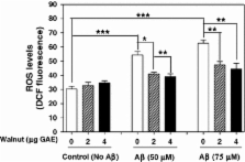
          Amyloid beta-protein (Aβ) is the major component of senile plaques and cerebrovascular amyloid deposits in individuals with Alzheimer’s disease. Aβ is known to increase free radical production in neuronal cells, leading to oxidative stress and cell death. Recently, considerable attention has been focused on dietary antioxidants that are able to scavenge reactive oxygen species (ROS), thereby offering protection against oxidative stress. Walnuts are rich in components that have anti-oxidant and anti-inflammatory properties. The inhibition of in vitro fibrillization of synthetic Aβ, and solubilization of preformed fibrillar Aβ by walnut extract was previously reported. The present study was designed to investigate whether walnut extract can protect against Aβ-induced oxidative damage and cytotoxicity. The effect of walnut extract on Aβ-induced cellular damage, ROS generation and apoptosis in PC12 pheochromocytoma cells was studied. Walnut extract reduced Aβ-mediated cell death assessed by MTT (3-(4,5-dimethylthiazol-2-yl)-2,5-diphenyltetrazolium bromide) reduction, and release of lactate dehydrogenase (membrane damage), DNA damage (apoptosis) and generation of ROS in a concentration-dependent manner. These results suggest that walnut extract can counteract Aβ-induced oxidative stress and associated cell death.

          Related collections

          Most cited references62

          • Record: found
          • Abstract: found
          • Article: not found

          Flavonoids as antioxidants.

          Flavonoids are phenolic substances isolated from a wide range of vascular plants, with over 8000 individual compounds known. They act in plants as antioxidants, antimicrobials, photoreceptors, visual attractors, feeding repellants, and for light screening. Many studies have suggested that flavonoids exhibit biological activities, including antiallergenic, antiviral, antiinflammatory, and vasodilating actions. However, most interest has been devoted to the antioxidant activity of flavonoids, which is due to their ability to reduce free radical formation and to scavenge free radicals. The capacity of flavonoids to act as antioxidants in vitro has been the subject of several studies in the past years, and important structure-activity relationships of the antioxidant activity have been established. The antioxidant efficacy of flavonoids in vivo is less documented, presumably because of the limited knowledge on their uptake in humans. Most ingested flavonoids are extensively degraded to various phenolic acids, some of which still possess a radical-scavenging ability. Both the absorbed flavonoids and their metabolites may display an in vivo antioxidant activity, which is evidenced experimentally by the increase of the plasma antioxidant status, the sparing effect on vitamin E of erythrocyte membranes and low-density lipoproteins, and the preservation of erythrocyte membrane polyunsaturated fatty acids. This review presents the current knowledge on structural aspects and in vitro antioxidant capacity of most common flavonoids as well as in vivo antioxidant activity and effects on endogenous antioxidants.
            Bookmark
            • Record: found
            • Abstract: found
            • Article: not found

            A specific amyloid-beta protein assembly in the brain impairs memory.

            Memory function often declines with age, and is believed to deteriorate initially because of changes in synaptic function rather than loss of neurons. Some individuals then go on to develop Alzheimer's disease with neurodegeneration. Here we use Tg2576 mice, which express a human amyloid-beta precursor protein (APP) variant linked to Alzheimer's disease, to investigate the cause of memory decline in the absence of neurodegeneration or amyloid-beta protein amyloidosis. Young Tg2576 mice ( 14 months old) form abundant neuritic plaques containing amyloid-beta (refs 3-6). We found that memory deficits in middle-aged Tg2576 mice are caused by the extracellular accumulation of a 56-kDa soluble amyloid-beta assembly, which we term Abeta*56 (Abeta star 56). Abeta*56 purified from the brains of impaired Tg2576 mice disrupts memory when administered to young rats. We propose that Abeta*56 impairs memory independently of plaques or neuronal loss, and may contribute to cognitive deficits associated with Alzheimer's disease.
              Bookmark
              • Record: found
              • Abstract: found
              • Article: not found

              Redox environment of the cell as viewed through the redox state of the glutathione disulfide/glutathione couple.

              Redox state is a term used widely in the research field of free radicals and oxidative stress. Unfortunately, it is used as a general term referring to relative changes that are not well defined or quantitated. In this review we provide a definition for the redox environment of biological fluids, cell organelles, cells, or tissue. We illustrate how the reduction potential of various redox couples can be estimated with the Nernst equation and show how pH and the concentrations of the species comprising different redox couples influence the reduction potential. We discuss how the redox state of the glutathione disulfide-glutathione couple (GSSG/2GSH) can serve as an important indicator of redox environment. There are many redox couples in a cell that work together to maintain the redox environment; the GSSG/2GSH couple is the most abundant redox couple in a cell. Changes of the half-cell reduction potential (E(hc)) of the GSSG/2GSH couple appear to correlate with the biological status of the cell: proliferation E(hc) approximately -240 mV; differentiation E(hc) approximately -200 mV; or apoptosis E(hc) approximately -170 mV. These estimates can be used to more fully understand the redox biochemistry that results from oxidative stress. These are the first steps toward a new quantitative biology, which hopefully will provide a rationale and understanding of the cellular mechanisms associated with cell growth and development, signaling, and reductive or oxidative stress.
                Bookmark

                Author and article information

                Contributors
                +1-718-494-5258 , +1-718-6987916 , abha.chauhan@opwdd.ny.gov
                Journal
                Neurochem Res
                Neurochemical Research
                Springer US (Boston )
                0364-3190
                1573-6903
                25 June 2011
                25 June 2011
                November 2011
                : 36
                : 11
                : 2096-2103
                Affiliations
                Department of Neurochemistry, NYS Institute for Basic Research in Developmental Disabilities, 1050 Forest Hill Road, Staten Island, NY 10314 USA
                Article
                533
                10.1007/s11064-011-0533-z
                3183245
                21706234
                9e4dc02e-ca45-442e-9eda-890ff13ddf40
                © The Author(s) 2011
                History
                : 10 June 2011
                Categories
                Original Paper
                Custom metadata
                © Springer Science+Business Media, LLC 2011

                Neurosciences
                apoptosis,oxidative stress,amyloid beta-protein,cytotoxicity,alzheimer’s disease,walnut

                Comments

                Comment on this article