91
views
0
recommends
+1 Recommend
0 collections
    0
    shares
      • Record: found
      • Abstract: found
      • Article: not found

      Differential innate immune response programs in neuronal subtypes determine susceptibility to infection in the brain by positive stranded RNA viruses

      research-article

      Read this article at

      ScienceOpenPublisherPMC
      Bookmark
          There is no author summary for this article yet. Authors can add summaries to their articles on ScienceOpen to make them more accessible to a non-specialist audience.

          Abstract

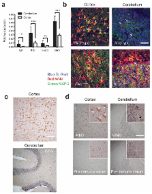
          Although susceptibility of neurons in the brain to microbial infection is a major determinant of clinical outcome, little is known about the molecular factors governing this. Here, we show that two types of neurons from distinct brain regions exhibited differential permissivity to replication of several positive-stranded RNA viruses. Granule cell neurons (GCN) of the cerebellum and cortical neurons (CN) from the cerebral cortex have unique innate immune programs that confer differential susceptibility to viral infection ex vivo and in vivo. By transducing CN with genes that were expressed more highly in GCN, we identified three interferon-stimulated genes (ISGs; Ifi27, Irg1, and Rsad2/Viperin) that mediated antiviral effects against different neurotropic viruses. Moreover, we found that the epigenetic state and microRNA-mediated regulation of ISGs correlates with enhanced antiviral response in GCN. Thus, neurons from evolutionarily distinct brain regions have unique innate immune signatures, which likely contribute to their relative permissiveness to infection.

          Related collections

          Most cited references41

          • Record: found
          • Abstract: found
          • Article: not found

          A MicroRNA feedback circuit in midbrain dopamine neurons.

          MicroRNAs (miRNAs) are evolutionarily conserved, 18- to 25-nucleotide, non-protein coding transcripts that posttranscriptionally regulate gene expression during development. miRNAs also occur in postmitotic cells, such as neurons in the mammalian central nervous system, but their function is less well characterized. We investigated the role of miRNAs in mammalian midbrain dopaminergic neurons (DNs). We identified a miRNA, miR-133b, that is specifically expressed in midbrain DNs and is deficient in midbrain tissue from patients with Parkinson's disease. miR-133b regulates the maturation and function of midbrain DNs within a negative feedback circuit that includes the paired-like homeodomain transcription factor Pitx3. We propose a role for this feedback circuit in the fine-tuning of dopaminergic behaviors such as locomotion.
            Bookmark
            • Record: found
            • Abstract: found
            • Article: not found

            A cAMP-response element binding protein-induced microRNA regulates neuronal morphogenesis.

            MicroRNAs (miRNAs) regulate cellular fate by controlling the stability or translation of mRNA transcripts. Although the spatial and temporal patterning of miRNA expression is tightly controlled, little is known about signals that induce their expression nor mechanisms of their transcriptional regulation. Furthermore, few miRNA targets have been validated experimentally. The miRNA, miR132, was identified through a genome-wide screen as a target of the transcription factor, cAMP-response element binding protein (CREB). miR132 is enriched in neurons and, like many neuronal CREB targets, is highly induced by neurotrophins. Expression of miR132 in cortical neurons induced neurite outgrowth. Conversely, inhibition of miR132 function attenuated neuronal outgrowth. We provide evidence that miR132 regulates neuronal morphogenesis by decreasing levels of the GTPase-activating protein, p250GAP. These data reveal that a CREB-regulated miRNA regulates neuronal morphogenesis by responding to extrinsic trophic cues.
              Bookmark
              • Record: found
              • Abstract: found
              • Article: not found

              Unbiased reconstruction of a mammalian transcriptional network mediating pathogen responses.

              Models of mammalian regulatory networks controlling gene expression have been inferred from genomic data but have largely not been validated. We present an unbiased strategy to systematically perturb candidate regulators and monitor cellular transcriptional responses. We applied this approach to derive regulatory networks that control the transcriptional response of mouse primary dendritic cells to pathogens. Our approach revealed the regulatory functions of 125 transcription factors, chromatin modifiers, and RNA binding proteins, which enabled the construction of a network model consisting of 24 core regulators and 76 fine-tuners that help to explain how pathogen-sensing pathways achieve specificity. This study establishes a broadly applicable, comprehensive, and unbiased approach to reveal the wiring and functions of a regulatory network controlling a major transcriptional response in primary mammalian cells.
                Bookmark

                Author and article information

                Journal
                9502015
                8791
                Nat Med
                Nat. Med.
                Nature medicine
                1078-8956
                1546-170X
                9 February 2013
                03 March 2013
                April 2013
                01 October 2013
                : 19
                : 4
                : 458-464
                Affiliations
                [1 ]Department of Molecular Microbiology, Washington University School of Medicine, St. Louis, Missouri 63110
                [2 ]Department of Medicine, Washington University School of Medicine, St. Louis, Missouri 63110
                [3 ]Department of Pathology and Immunology, Washington University School of Medicine, St. Louis, Missouri 63110
                [4 ]Department of Microbiology, University of Washington School of Medicine, Seattle, WA 98195-7650
                [5 ]Department of Immunology, University of Washington School of Medicine, Seattle, WA 98195-7650
                Author notes
                Corresponding author: Michael S. Diamond, M.D., Ph.D., Departments of Medicine, Molecular Microbiology and Pathology & Immunology, Washington University School of Medicine, 660 South Euclid Avenue, Box 8051, St Louis. Missouri 63110. Tel: 314-362-2842. Fax: 314-362-9230. diamond@ 123456borcim.wustl.edu
                Article
                NIHMS439947
                10.1038/nm.3108
                3618596
                23455712
                9e321005-8586-4dd3-9673-13fee62e3230

                Users may view, print, copy, download and text and data- mine the content in such documents, for the purposes of academic research, subject always to the full Conditions of use: http://www.nature.com/authors/editorial_policies/license.html#terms

                History
                Funding
                Funded by: National Institute of Allergy and Infectious Diseases Extramural Activities : NIAID
                Award ID: U54 AI081680 || AI
                Funded by: National Institute of Allergy and Infectious Diseases Extramural Activities : NIAID
                Award ID: U19 AI083019 || AI
                Funded by: National Institute of Allergy and Infectious Diseases Extramural Activities : NIAID
                Award ID: R01 AI074973 || AI
                Categories
                Article

                Medicine
                Medicine

                Comments

                Comment on this article