27
views
0
recommends
+1 Recommend
0 collections
    0
    shares
      • Record: found
      • Abstract: found
      • Article: found
      Is Open Access

      Inter- and intratumoral heterogeneity of BCL2 correlates with IgH expression and prognosis in follicular lymphoma

      research-article

      Read this article at

      Bookmark
          There is no author summary for this article yet. Authors can add summaries to their articles on ScienceOpen to make them more accessible to a non-specialist audience.

          Abstract

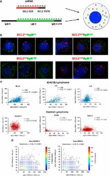
          Most follicular lymphomas (FLs) are genetically defined by the t(14;18)(q32;q21) translocation that juxtaposes the BCL2 gene to the immunoglobulin heavy chain (IgH) 3' regulatory regions (IgH-3'RRs). Despite this recurrent translocation, FL cases are heterogeneous in terms of intratumoral clonal diversity for acquired mutations and variations in the tumor microenvironment. Here we describe an additional mechanism that contributes to inter- and intratumoral heterogeneity in FLs. By applying a novel single-molecule RNA fluorescence-based in situ hybridization (FISH) technique to detect mRNA molecules of BCL2 and IgH in single cells, we found marked heterogeneity in the number of BCL2 mRNA transcripts within individual lymphoma cells. Moreover, BCL2 mRNA molecules correlated with IgH mRNA molecules in individual cells both in t(14;18) lymphoma cell lines and in patient samples. Consistently, a strong correlation between BCL2 and IgH protein levels was found in a series of 205 primary FL cases by flow cytometry and immunohistochemistry. Inter- and intratumoral heterogeneity of BCL2 expression determined resistance to drugs commonly used in FL treatment and affected overall survival of FL patients. These data demonstrate that BCL2 and IgH expressions are heterogeneous and coregulated in t(14;18)-translocated cells, and determine the response to therapy in FL patients.

          Related collections

          Most cited references39

          • Record: found
          • Abstract: found
          • Article: not found

          Follicular lymphoma international prognostic index 2: a new prognostic index for follicular lymphoma developed by the international follicular lymphoma prognostic factor project.

          The aim of the F2 study was to verify whether a prospective collection of data would enable the development of a more accurate prognostic index for follicular lymphoma (FL) by using parameters which could not be retrospectively studied before, and by choosing progression-free survival (PFS) as principal end point. Between January 2003 and May 2005, 1,093 patients with a newly diagnosed FL were registered and 942 individuals receiving antilymphoma therapy were selected as the study population. The variables we used for score definition were selected by means of bootstrap resampling procedures on 832 patients with complete data. Procedures to select the model that would minimize errors were also performed. After a median follow-up of 38 months, 261 events for PFS evaluation were recorded. beta2-microglobulin higher than the upper limit of normal, longest diameter of the largest involved node longer than 6 cm, bone marrow involvement, hemoglobin level lower than 12 g/dL, and age older than 60 years were factors independently predictive for PFS. Using these variables, a prognostic model was devised to identify three groups at different levels of risk. The 3-year PFS rate was 91%, 69%, and 51% for patients at low, intermediate, and high risk, respectively (log-rank = 64.6; P < .00001). The 3-year survival rate was 99%, 96%, and 84% for patients at low, intermediate, and high risk, respectively (P < .0001). Follicular Lymphoma International Prognostic Index 2 is a simple prognostic index based on easily available clinical data and may represent a promising new tool for the identification of patients with FL at different risk in the era of immunochemotherapy.
            Bookmark
            • Record: found
            • Abstract: found
            • Article: not found

            Involvement of the bcl-2 gene in human follicular lymphoma.

            Recombinant DNA probes were cloned for the areas flanking the breakpoint on chromosome 18 in cells from a patient with acute lymphocytic leukemia of the B-cell type; cells of this line carry the t(14;18) chromosomal translocation. Two of the probes detected DNA rearrangements in approximately 60 percent of the cases of follicular lymphoma screened. In follicular lymphoma, most of the breakpoints in band q21 of chromosome 18 were clustered within a short stretch of DNA, approximately 2.1 kilobases in length. Chromosome 18-specific DNA probes for the areas flanking the breakpoints also detected RNA transcripts 6 kilobases in length in various cell types. The gene coding for these transcript (the bcl-2 gene) seems to be interrupted in most cases of follicular lymphomas carrying the t(14;18) chromosomal translocation.
              Bookmark
              • Record: found
              • Abstract: found
              • Article: not found

              Hierarchy in somatic mutations arising during genomic evolution and progression of follicular lymphoma.

              Follicular lymphoma (FL) is currently incurable using conventional chemotherapy or immunotherapy regimes, compelling new strategies. Advances in high-throughput sequencing technologies that can reveal oncogenic pathways have stimulated interest in tailoring therapies toward actionable somatic mutations. However, for mutation-directed therapies to be most effective, the mutations must be uniformly present in evolved tumor cells as well as in the self-renewing tumor-cell precursors. Here, we show striking intratumoral clonal diversity within FL tumors in the representation of mutations in the majority of genes as revealed by whole exome sequencing of subpopulations. This diversity captures a clonal hierarchy, resolved using immunoglobulin somatic mutations and IGH-BCL2 translocations as a frame of reference and by comparing diagnosis and relapse tumor pairs, allowing us to distinguish early versus late genetic eventsduring lymphomagenesis. We provide evidence that IGH-BCL2 translocations and CREBBP mutations are early events, whereas MLL2 and TNFRSF14 mutations probably represent late events during disease evolution. These observations provide insight into which of the genetic lesions represent suitable candidates for targeted therapies.
                Bookmark

                Author and article information

                Journal
                Blood Cancer J
                Blood Cancer J
                Blood Cancer Journal
                Nature Publishing Group
                2044-5385
                October 2014
                10 October 2014
                1 October 2014
                : 4
                : 10
                : e249
                Affiliations
                [1 ]Department of Molecular Biotechnology and Health Sciences, University of Torino , Torino, Italy
                [2 ]Center for Experimental Medicine and Clinical Studies (CERMS), University of Torino , Torino, Italy
                [3 ]Department of Medical Sciences, University of Torino , Torino, Italy
                [4 ]Department of Clinical and Biological Sciences, University of Torino , Torino, Italy
                [5 ]ASO San Giovanni Battista Hospital , Turin, Italy
                [6 ]Departments of Physics, Massachusetts Institute of Technology , Cambridge, MA, USA
                [7 ]Department of Biology, Massachusetts Institute of Technology , Cambridge, MA, USA
                [8 ]Koch Institute for Integrative Cancer Research, Massachusetts Institute of Technology , Cambridge, MA, USA
                [9 ]Hubrecht Institute–KNAW (Royal Netherlands Academy of Arts and Sciences) , Utrecht, The Netherlands
                [10 ]University Medical Center Utrecht , Utrecht, The Netherlands
                [11 ]Department of Pathology, Children's Hospital , Boston, MA, USA
                [12 ]Harvard Medical School, Boston, MA, USA
                Author notes
                [* ]Hubrecht Institute–KNAW (Royal Netherlands Academy of Arts and Sciences), University Medical Center Utrecht , Utrecht, The Netherlands E-mail: n.crosetto@ 123456hubrecht.eu
                [* ]Department of Pathology, Children's Hospital Boston and Harvard Medical School, Enders 1116.1 , 320 Longwood Avenue, Boston, MA 02115, USA. E-mail: roberto.chiarle@ 123456childrens.harvard.edu
                [13]

                These authors contributed equally to this work.

                Article
                bcj201467
                10.1038/bcj.2014.67
                4220646
                25303368
                9e27f649-c06e-4b73-9120-2fb593941b85
                Copyright © 2014 Macmillan Publishers Limited

                This work is licensed under a Creative Commons Attribution-NonCommercial-ShareAlike 4.0 International License. The images or other third party material in this article are included in the article's Creative Commons license, unless indicated otherwise in the credit line; if the material is not included under the Creative Commons license, users will need to obtain permission from the license holder to reproduce the material. To view a copy of this license, visit http://creativecommons.org/licenses/by-nc-sa/4.0/

                History
                : 13 August 2014
                : 20 August 2014
                Categories
                Original Article

                Oncology & Radiotherapy
                Oncology & Radiotherapy

                Comments

                Comment on this article