8
views
0
recommends
+1 Recommend
0 collections
    0
    shares
      • Record: found
      • Abstract: found
      • Article: found
      Is Open Access

      Alpha Ketoglutarate Exerts In Vitro Anti-Osteosarcoma Effects through Inhibition of Cell Proliferation, Induction of Apoptosis via the JNK and Caspase 9-Dependent Mechanism, and Suppression of TGF-β and VEGF Production and Metastatic Potential of Cells

      research-article

      Read this article at

      Bookmark
          There is no author summary for this article yet. Authors can add summaries to their articles on ScienceOpen to make them more accessible to a non-specialist audience.

          Abstract

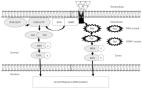
          Osteosarcoma (OS) is the most common type of primary bone tumor. Currently, there are limited treatment options for metastatic OS. Alpha-ketoglutarate (AKG), i.e., a multifunctional intermediate of the Krebs cycle, is one of the central metabolic regulators of tumor fate and plays an important role in cancerogenesis and tumor progression. There is growing evidence suggesting that AKG may represent a novel adjuvant therapeutic opportunity in anti-cancer therapy. The present study was intended to check whether supplementation of Saos-2 and HOS osteosarcoma cell lines (harboring a TP53 mutation) with exogenous AKG exerted an anti-cancer effect. The results revealed that AKG inhibited the proliferation of both OS cell lines in a concentration-dependent manner. As evidenced by flow cytometry, AKG blocked cell cycle progression at the G 1 stage in both cell lines, which was accompanied by a decreased level of cyclin D1 in HOS and increased expression of p21 Waf1/Cip1 protein in Saos-2 cells (evaluated with the ELISA method). Moreover, AKG induced apoptotic cell death and caspase-3 activation in both OS cell lines (determined by cytometric analysis). Both the immunoblotting and cytometric analysis revealed that the AKG-induced apoptosis proceeded predominantly through activation of an intrinsic caspase 9-dependent apoptotic pathway and an increased Bax/Bcl-2 ratio. The apoptotic process in the AKG-treated cells was mediated via c-Jun N-terminal protein kinase (JNK) activation, as the specific inhibitor of this kinase partially rescued the cells from apoptotic death. In addition, the AKG treatment led to reduced activation of extracellular signal-regulated kinase (ERK1/2) and significant inhibition of cell migration and invasion in vitro concomitantly with decreased production of pro-metastatic transforming growth factor β (TGF-β) and pro-angiogenic vascular endothelial growth factor (VEGF) in both OS cell lines suggesting the anti-metastatic potential of this compound. In conclusion, we showed the anti-osteosarcoma potential of AKG and provided a rationale for a further study of the possible application of AKG in OS therapy.

          Related collections

          Most cited references73

          • Record: found
          • Abstract: found
          • Article: not found

          Activation of apoptosis signalling pathways by reactive oxygen species.

          Reactive oxygen species (ROS) are short-lived and highly reactive molecules. The generation of ROS in cells exists in equilibrium with a variety of antioxidant defences. At low to modest doses, ROS are considered to be essential for regulation of normal physiological functions involved in development such as cell cycle progression and proliferation, differentiation, migration and cell death. ROS also play an important role in the immune system, maintenance of the redox balance and have been implicated in activation of various cellular signalling pathways. Excess cellular levels of ROS cause damage to proteins, nucleic acids, lipids, membranes and organelles, which can lead to activation of cell death processes such as apoptosis. Apoptosis is a highly regulated process that is essential for the development and survival of multicellular organisms. These organisms often need to discard cells that are superfluous or potentially harmful, having accumulated mutations or become infected by pathogens. Apoptosis features a characteristic set of morphological and biochemical features whereby cells undergo a cascade of self-destruction. Thus, proper regulation of apoptosis is essential for maintaining normal cellular homeostasis. ROS play a central role in cell signalling as well as in regulation of the main pathways of apoptosis mediated by mitochondria, death receptors and the endoplasmic reticulum (ER). This review focuses on current understanding of the role of ROS in each of these three main pathways of apoptosis. The role of ROS in the complex interplay and crosstalk between these different signalling pathways remains to be further unravelled during the coming years.
            Bookmark
            • Record: found
            • Abstract: found
            • Article: found
            Is Open Access

            ERK/MAPK signalling pathway and tumorigenesis

            Mitogen-activated protein kinase (MAPK) cascades are key signalling pathways that regulate a wide variety of cellular processes, including proliferation, differentiation, apoptosis and stress responses. The MAPK pathway includes three main kinases, MAPK kinase kinase, MAPK kinase and MAPK, which activate and phosphorylate downstream proteins. The extracellular signal-regulated kinases ERK1 and ERK2 are evolutionarily conserved, ubiquitous serine-threonine kinases that regulate cellular signalling under both normal and pathological conditions. ERK expression is critical for development and their hyperactivation plays a major role in cancer development and progression. The Ras/Raf/MAPK (MEK)/ERK pathway is the most important signalling cascade among all MAPK signal transduction pathways, and plays a crucial role in the survival and development of tumour cells. The present review discusses recent studies on Ras and ERK pathway members. With respect to processes downstream of ERK activation, the role of ERK in tumour proliferation, invasion and metastasis is highlighted, and the role of the ERK/MAPK signalling pathway in tumour extracellular matrix degradation and tumour angiogenesis is emphasised.
              Bookmark
              • Record: found
              • Abstract: found
              • Article: not found

              Oncometabolite 2-hydroxyglutarate is a competitive inhibitor of α-ketoglutarate-dependent dioxygenases.

              IDH1 and IDH2 mutations occur frequently in gliomas and acute myeloid leukemia, leading to simultaneous loss and gain of activities in the production of α-ketoglutarate (α-KG) and 2-hydroxyglutarate (2-HG), respectively. Here we demonstrate that 2-HG is a competitive inhibitor of multiple α-KG-dependent dioxygenases, including histone demethylases and the TET family of 5-methlycytosine (5mC) hydroxylases. 2-HG occupies the same space as α-KG does in the active site of histone demethylases. Ectopic expression of tumor-derived IDH1 and IDH2 mutants inhibits histone demethylation and 5mC hydroxylation. In glioma, IDH1 mutations are associated with increased histone methylation and decreased 5-hydroxylmethylcytosine (5hmC). Hence, tumor-derived IDH1 and IDH2 mutations reduce α-KG and accumulate an α-KG antagonist, 2-HG, leading to genome-wide histone and DNA methylation alterations. Copyright © 2011 Elsevier Inc. All rights reserved.
                Bookmark

                Author and article information

                Journal
                Int J Mol Sci
                Int J Mol Sci
                ijms
                International Journal of Molecular Sciences
                MDPI
                1422-0067
                10 December 2020
                December 2020
                : 21
                : 24
                : 9406
                Affiliations
                [1 ]Department of Virology and Immunology, Maria Curie-Sklodowska University, Akademicka 19, 20-033 Lublin, Poland; katarzyna.kalawaj@ 123456op.pl (K.K.); magdalena.mizerska-dudka@ 123456poczta.umcs.lublin.pl (M.M.-K.); zurekaleksandra@ 123456wp.pl (A.Ż.); kandem@ 123456poczta.umcs.lublin.pl (M.K.-S.)
                [2 ]Department of Cell Biology, Maria Curie-Sklodowska University, Akademicka 19, 20-033 Lublin, Poland; adrianna.slawinska-brych@ 123456poczta.umcs.lublin.pl
                [3 ]Chair and Department of Clinical Immunology, Medical University of Lublin, Chodźki 4a, 20-093 Lublin, Poland; agnieszka.bojarska-junak@ 123456umlub.pl
                Author notes
                Author information
                https://orcid.org/0000-0003-2364-6181
                https://orcid.org/0000-0003-2340-9442
                https://orcid.org/0000-0002-0920-3192
                Article
                ijms-21-09406
                10.3390/ijms21249406
                7763003
                33321940
                9e0e32e6-06d8-428c-9e2a-500b5da0f0ae
                © 2020 by the authors.

                Licensee MDPI, Basel, Switzerland. This article is an open access article distributed under the terms and conditions of the Creative Commons Attribution (CC BY) license ( http://creativecommons.org/licenses/by/4.0/).

                History
                : 10 November 2020
                : 08 December 2020
                Categories
                Article

                Molecular biology
                alpha-ketoglutarate,cell cycle,apoptosis,jnk,cell migration,cell invasion,tgf-β,vegf
                Molecular biology
                alpha-ketoglutarate, cell cycle, apoptosis, jnk, cell migration, cell invasion, tgf-β, vegf

                Comments

                Comment on this article