16
views
0
recommends
+1 Recommend
0 collections
    0
    shares
      • Record: found
      • Abstract: found
      • Article: found
      Is Open Access

      Mapping the interactions of the single-stranded DNA binding protein of bacteriophage T4 (gp32) with DNA lattices at single nucleotide resolution: polynucleotide binding and cooperativity

      research-article

      Read this article at

      Bookmark
          There is no author summary for this article yet. Authors can add summaries to their articles on ScienceOpen to make them more accessible to a non-specialist audience.

          Abstract

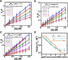
          We here use our site-specific base analog mapping approach to study the interactions and binding equilibria of cooperatively-bound clusters of the single-stranded DNA binding protein (gp32) of the T4 DNA replication complex with longer ssDNA (and dsDNA) lattices. We show that in cooperatively bound clusters the binding free energy appears to be equi-partitioned between the gp32 monomers of the cluster, so that all bind to the ssDNA lattice with comparable affinity, but also that the outer domains of the gp32 monomers at the ends of the cluster can fluctuate on and off the lattice and that the clusters of gp32 monomers can slide along the ssDNA. We also show that at very low binding densities gp32 monomers bind to the ssDNA lattice at random, but that cooperatively bound gp32 clusters bind preferentially at the 5′-end of the ssDNA lattice. We use these results and the gp32 monomer-binding results of the companion paper to propose a detailed model for how gp32 might bind to and interact with ssDNA lattices in its various binding modes, and also consider how these clusters might interact with other components of the T4 DNA replication complex.

          Related collections

          Most cited references26

          • Record: found
          • Abstract: not found
          • Article: not found

          Theoretical aspects of DNA-protein interactions: co-operative and non-co-operative binding of large ligands to a one-dimensional homogeneous lattice.

            Bookmark
            • Record: found
            • Abstract: found
            • Article: not found

            Crystal structure of a replication fork single-stranded DNA binding protein (T4 gp32) complexed to DNA.

            The single-stranded DNA (ssDNA) binding protein gp32 from bacteriophage T4 is essential for T4 DNA replication, recombination and repair. In vivo gp32 binds ssDNA as the replication fork advances and stimulates replisome processivity and accuracy by a factor of several hundred. Gp32 binding affects nearly every major aspect of DNA metabolism. Among its important functions are: (1) configuring ssDNA templates for efficient use by the replisome including DNA polymerase; (2) melting out adventitious secondary structures; (3) protecting exposed ssDNA from nucleases; and (4) facilitating homologous recombination by binding ssDNA during strand displacement. We have determined the crystal structure of the gp32 DNA binding domain complexed to ssDNA at 2.2 A resolution. The ssDNA binding cleft comprises regions from three structural subdomains and includes a positively charged surface that runs parallel to a series of hydrophobic pockets formed by clusters of aromatic side chains. Although only weak electron density is seen for the ssDNA, it indicates that the phosphate backbone contacts an electropositive cleft of the protein, placing the bases in contact with the hydrophobic pockets. The DNA mobility implied by the weak electron density may reflect the role of gp32 as a sequence-independent ssDNA chaperone allowing the largely unstructured ssDNA to slide freely through the cleft.
              Bookmark
              • Record: found
              • Abstract: found
              • Article: not found

              Spectroscopic studies of position-specific DNA "breathing" fluctuations at replication forks and primer-template junctions.

              Junctions between ssDNA and dsDNA sequences are important in many cellular processes, including DNA replication, transcription, recombination, and repair. Significant transient conformational fluctuations ("DNA breathing") can occur at these ssDNA-dsDNA junctions. The involvement of such breathing in the mechanisms of macromolecular complexes that operate at these loci is not well understood, in part because these fluctuations have been difficult to measure in a position-specific manner. To address this issue we constructed forked or primer-template DNA constructs with 1 or 2 adjacent 2-aminopurine (2-AP) nucleotide residues (adenine analogues) placed at specific positions on both sides of the ssDNA-dsDNA junction. Unlike canonical DNA bases, 2-AP absorbs, fluoresces, and displays CD spectra at wavelengths >300 nm, where other nucleic acid and protein components are transparent. We used CD and fluorescence spectra and acrylamide quenching of these probes to monitor the extent and nature of DNA breathing of A-T base pairs at specific positions around the ssDNA-dsDNA junction. As expected, spectroscopically measurable unwinding penetrates approximately 2 bp into the duplex region of these junctions under physiological conditions for the constructs examined. Surprisingly, we found that 2-AP bases at ssDNA sites directly adjacent to ssDNA-dsDNA junctions are significantly more unstacked than those at more distant ssDNA positions. These local and transient DNA conformations on both sides of ssDNA-dsDNA junctions may serve as specific interaction targets for enzymes that manipulate DNA in the processes of gene expression.
                Bookmark

                Author and article information

                Journal
                Nucleic Acids Res
                Nucleic Acids Res
                nar
                nar
                Nucleic Acids Research
                Oxford University Press
                0305-1048
                1362-4962
                30 October 2015
                14 August 2015
                14 August 2015
                : 43
                : 19
                : 9291-9305
                Affiliations
                Institute of Molecular Biology and Department of Chemistry, University of Oregon, Eugene, OR 97403-1229, USA
                Author notes
                [* ]To whom correspondence should be addressed. Tel: +1 541 346 5151; Fax: +1 541 346 5891; Email: petevh@ 123456molbio.uoregon.edu
                Article
                10.1093/nar/gkv818
                4627071
                26275774
                9ddcd458-c9e1-41e8-9bf4-f31babdfdeae
                © The Author(s) 2015. Published by Oxford University Press on behalf of Nucleic Acids Research.

                This is an Open Access article distributed under the terms of the Creative Commons Attribution License ( http://creativecommons.org/licenses/by-nc/4.0/), which permits non-commercial re-use, distribution, and reproduction in any medium, provided the original work is properly cited. For commercial re-use, please contact journals.permissions@ 123456oup.com

                History
                : 31 July 2015
                : 13 July 2015
                : 14 March 2015
                Page count
                Pages: 15
                Categories
                16
                Genome Integrity, Repair and Replication
                Custom metadata
                30 October 2015

                Genetics
                Genetics

                Comments

                Comment on this article