6
views
0
recommends
+1 Recommend
0 collections
    0
    shares
      • Record: found
      • Abstract: found
      • Article: found
      Is Open Access

      Feedback control of PLK1 by Apolo1 ensures accurate chromosome segregation

      research-article

      Read this article at

      Bookmark
          There is no author summary for this article yet. Authors can add summaries to their articles on ScienceOpen to make them more accessible to a non-specialist audience.

          SUMMARY

          Stable transmission of genetic material during cell division requires accurate chromosome segregation. PLK1 dynamics at kinetochores control establishment of correct kinetochore-microtubule attachments and subsequent silencing of the spindle checkpoint. However, the regulatory mechanism responsible for PLK1 activity in prometaphase has not yet been affirmatively identified. Here we identify Apolo1, which tunes PLK1 activity for accurate kinetochore-microtubule attachments. Apolo1 localizes to kinetochores during early mitosis, and suppression of Apolo1 results in misaligned chromosomes. Using the fluorescence resonance energy transfer (FRET)-based PLK1 activity reporter, we found that Apolo1 sustains PLK1 kinase activity at kinetochores for accurate attachment during prometaphase. Apolo1 is a cognate substrate of PLK1, and the phosphorylation enables PP1γ to inactivate PLK1 by dephosphorylation. Mechanistically, Apolo1 constitutes a bridge between kinase and phosphatase, which governs PLK1 activity in prometaphase. These findings define a previously uncharacterized feedback loop by which Apolo1 provides fine-tuning for PLK1 to guide chromosome segregation in mitosis.

          Graphical abstract
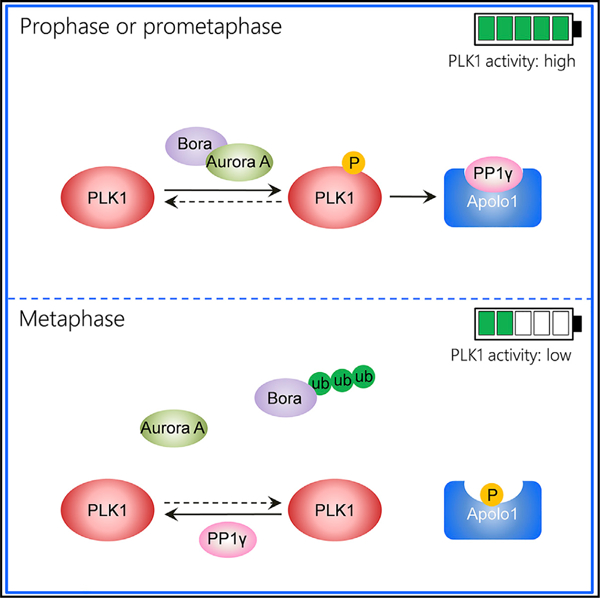
          In brief

          Xu et al. identify Apolo1, which governs PLK1 activity and promotes faithful chromosome segregation in prometaphase by bridging kinase and phosphatase activities.

          Related collections

          Most cited references50

          • Record: found
          • Abstract: found
          • Article: not found

          A gene-coexpression network for global discovery of conserved genetic modules.

          To elucidate gene function on a global scale, we identified pairs of genes that are coexpressed over 3182 DNA microarrays from humans, flies, worms, and yeast. We found 22,163 such coexpression relationships, each of which has been conserved across evolution. This conservation implies that the coexpression of these gene pairs confers a selective advantage and therefore that these genes are functionally related. Many of these relationships provide strong evidence for the involvement of new genes in core biological functions such as the cell cycle, secretion, and protein expression. We experimentally confirmed the predictions implied by some of these links and identified cell proliferation functions for several genes. By assembling these links into a gene-coexpression network, we found several components that were animal-specific as well as interrelationships between newly evolved and ancient modules.
            Bookmark
            • Record: found
            • Abstract: found
            • Article: not found

            Proteogenomics connects somatic mutations to signaling in breast cancer

            Summary Somatic mutations have been extensively characterized in breast cancer, but the effects of these genetic alterations on the proteomic landscape remain poorly understood. We describe quantitative mass spectrometry-based proteomic and phosphoproteomic analyses of 105 genomically annotated breast cancers of which 77 provided high-quality data. Integrated analyses allowed insights into the somatic cancer genome including the consequences of chromosomal loss, such as the 5q deletion characteristic of basal-like breast cancer. The 5q trans effects were interrogated against the Library of Integrated Network-based Cellular Signatures, thereby connecting CETN3 and SKP1 loss to elevated expression of EGFR, and SKP1 loss also to increased SRC. Global proteomic data confirmed a stromal-enriched group in addition to basal and luminal clusters and pathway analysis of the phosphoproteome identified a G Protein-coupled receptor cluster that was not readily identified at the mRNA level. Besides ERBB2, other amplicon-associated, highly phosphorylated kinases were identified, including CDK12, PAK1, PTK2, RIPK2 and TLK2. We demonstrate that proteogenomic analysis of breast cancer elucidates functional consequences of somatic mutations, narrows candidate nominations for driver genes within large deletions and amplified regions, and identifies therapeutic targets.
              Bookmark
              • Record: found
              • Abstract: found
              • Article: not found

              Large-scale meta-analysis of cancer microarray data identifies common transcriptional profiles of neoplastic transformation and progression.

              Many studies have used DNA microarrays to identify the gene expression signatures of human cancer, yet the critical features of these often unmanageably large signatures remain elusive. To address this, we developed a statistical method, comparative metaprofiling, which identifies and assesses the intersection of multiple gene expression signatures from a diverse collection of microarray data sets. We collected and analyzed 40 published cancer microarray data sets, comprising 38 million gene expression measurements from >3,700 cancer samples. From this, we characterized a common transcriptional profile that is universally activated in most cancer types relative to the normal tissues from which they arose, likely reflecting essential transcriptional features of neoplastic transformation. In addition, we characterized a transcriptional profile that is commonly activated in various types of undifferentiated cancer, suggesting common molecular mechanisms by which cancer cells progress and avoid differentiation. Finally, we validated these transcriptional profiles on independent data sets.
                Bookmark

                Author and article information

                Journal
                101573691
                39703
                Cell Rep
                Cell Rep
                Cell reports
                2211-1247
                22 July 2021
                13 July 2021
                12 August 2021
                : 36
                : 2
                : 109343
                Affiliations
                [1 ]MOE Key Laboratory for Membraneless Organelles and Cellular Dynamics, University of Science & Technology of China, Hefei 230027, China
                [2 ]Keck Center for Molecular Imaging, Morehouse School of Medicine, Atlanta, GA 30310, USA
                [3 ]Anhui Key Laboratory for Chemical Biology, Hefei National Center for Physical Sciences at Microscale, Hefei 230027, China
                [4 ]Department of Chemistry, Southern University of Science and Technology, Shenzhen 518055, China
                [5 ]National Chromatographic Research and Analysis Center, Dalian 116023, China
                [6 ]Max Planck Institute for Molecular Cell Biology and Genetics, Pfotenhauerstrasse 108, 01307 Dresden, Germany
                [7 ]Shanghai Institute of Biochemistry and Cell Biology, Chinese Academy of Sciences, Shanghai 200031, China
                [8 ]These authors contributed equally
                [9 ]Lead contact
                Author notes

                AUTHOR CONTRIBUTIONS

                L.X. and X. Yao conceived the project. L.X. and M.A. designed and performed most biochemical experiments, cell biological characterization, and data analyses. X. Yuan and R.T. performed in vitro phosphorylation site identification and B.S. performed in vivo Apolo1 binding protein identification using mass spectrometric analyses. L.X., Y.G., L.C., and W.D. performed the FRET sensor assay, and D.L. constructed the PLK1 sensor. M.M. and F.G. conducted immunofluorescence analyses. A.H., I.P., Z.D., H.D., J.L., W.P., D.W., L.L., and H.G. contributed reagents. L.X., M.A., X. Yuan, Z.D., D.L., X.L., and X. Yao wrote the manuscript, and all authors have read and approved the manuscript.

                Article
                NIHMS1724377
                10.1016/j.celrep.2021.109343
                8358895
                34260926
                9ce3865b-2c9d-4db4-b2e9-281af896b4e7

                This is an open access article under the CC BY-NC-ND license ( http://creativecommons.org/licenses/by-nc-nd/4.0/).

                History
                Categories
                Article

                Cell biology
                Cell biology

                Comments

                Comment on this article