23
views
0
recommends
+1 Recommend
0 collections
    0
    shares
      • Record: found
      • Abstract: found
      • Article: found
      Is Open Access

      Spontaneous Pneumomediastinum, Pneumopericardium, and Pneumothorax with Respiratory Failure in a Patient with AIDS and Pneumocystis jirovecii Pneumonia

      case-report

      Read this article at

      Bookmark
          There is no author summary for this article yet. Authors can add summaries to their articles on ScienceOpen to make them more accessible to a non-specialist audience.

          Abstract

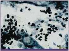
          Spontaneous pneumothorax occurs in up to 35% of patients with Pneumocystis jirovecii pneumonia. However, spontaneous pneumomediastinum and pneumopericardium are uncommon complications in patients infected with human immunodeficiency virus, with no reported incidence rates, even among patients with acquired immunodeficiency syndrome (AIDS) and P. jirovecii pneumonia. We report a case of spontaneous pneumomediastinum, pneumopericardium, and pneumothorax with respiratory failure during treatment of P. jirovecii pneumonia in a patient with AIDS; the P. jirovecii infection was confirmed by performing methenamine silver staining of bronchoalveolar lavage specimens. This case suggests that spontaneous pneumomediastinum and pneumopericardium should be considered in patients with AIDS and P. jirovecii pneumonia.

          Related collections

          Most cited references19

          • Record: found
          • Abstract: found
          • Article: found
          Is Open Access

          A New Name for Pneumocystis from Humans and New Perspectives on the Host-Pathogen Relationship

          The disease known as Pneumocystis carinii pneumonia (PCP) is a major cause of illness and death in persons with impaired immune systems. While the genus Pneumocystis has been known to science for nearly a century, understanding of its members remained rudimentary until DNA analysis showed its extensive diversity. Pneumocystis organisms from different host species have very different DNA sequences, indicating multiple species. In recognition of its genetic and functional distinctness, the organism that causes human PCP is now named Pneumocystis jiroveci Frenkel 1999. Changing the organism’s name does not preclude the use of the acronym PCP because it can be read “ Pneumocystis pneumonia.” DNA varies in samples of P. jiroveci, a feature that allows reexamination of the relationships between host and pathogen. Instead of lifelong latency, transient colonization may be the rule.
            Bookmark
            • Record: found
            • Abstract: found
            • Article: not found

            Spontaneous pneumomediastinum: analysis of 62 consecutive adult patients.

            To clarify the clinical presentation and course of patients with spontaneous pneumomediastinum (SP) and to determine the usefulness of diagnostic testing in these patients. We conducted a retrospective review of 62 consecutive adult patients (age >or=18 years) diagnosed as having SP during an 11-year period from July 1, 1997, to June 30, 2008. The study cohort included 41 men and 21 women (median age, 30 years; interquartile range, 20-69 years). Among the 62 study patients, the most common presenting symptoms were chest pain (39 patients [63%]), cough (28 [45%]), and dyspnea (27 [44%]). Preexisting lung diseases were identified in 27 patients (44%) and included interstitial lung disease, asthma, lung malignancies, bronchiolitis obliterans syndrome, chronic obstructive pulmonary disease, bronchiectasis, and cystic lung lesions. The initial diagnosis of SP was achieved by chest radiography in 52 patients (84%); the remaining cases were diagnosed by computed tomography. Forty-seven patients (76%) were hospitalized for a median duration of 2.5 days. Additional diagnostic procedures were performed in 27 patients (44%) and included contrast esophagography, bronchoscopy, and esophagogastroduodenoscopy; however, they did not yield a pathologic cause in any patient. Pneumothorax was identified in 20 patients (32%), but less than one-third of these patients underwent chest tube thoracostomy. No episodes of mediastinitis or sepsis occurred. Recurrence of SP was seen in 1 patient, and thoracoscopic surgery was performed in 1 patient for persistent air leak (pneumothorax). Spontaneous pneumomediastinum was associated with a relatively benign clinical course; however, pneumothorax was seen in 32% of cases. Diagnostic testing to determine a pathologic cause yielded little clinically relevant information in these patients.
              Bookmark
              • Record: found
              • Abstract: found
              • Article: not found

              Pneumothorax.

              Spontaneous pneumothoraces can occur without obvious underlying lung disease (primary) or in patients with known underlying lung disease (secondary). Management guidelines for spontaneous pneumothorax have been published by major professional organizations, but awareness and application among clinicians seems poor. First episodes of primary spontaneous pneumothorax can be managed with observation if the pneumothorax is small. If the pneumothorax is large or if the patient is symptomatic, manual aspiration via a small catheter or insertion of a small-bore catheter coupled to a Heimlich valve or water-seal device, should be performed. In general, definitive measures to prevent recurrence are recommended after the first recurrence of the pneumothorax, and can be achieved by medical (e.g. talc) or surgical (video-assisted thoracic surgery) pleurodesis. Secondary pneumothoraces should be treated with chest tube drainage followed by pleurodesis after the first episode to minimize any risk of recurrence. Traumatic pneumothoraces may be occult (not seen on an initial CXR) or non-occult. The majority are treated by placement of a chest tube. Selected patients may be treated conservatively, with approximately 10% of these patients eventually requiring chest tube placement. Iatrogenic pneumothoraces have a myriad of causes with transthoracic lung needle biopsy being most common. Transthoracic needle biopsy-related pneumothoraces have CT findings that can predict their occurrence and the need for chest tube placement. Iatrogenic pneumothoraces, regardless of cause, may be managed by observation or small bore chest tube placement, depending upon patient stability and the size of the pneumothorax.
                Bookmark

                Author and article information

                Journal
                Infect Chemother
                Infect Chemother
                IC
                Infection & Chemotherapy
                The Korean Society of Infectious Diseases and Korean Society for Chemotherapy
                2093-2340
                2092-6448
                September 2014
                24 September 2014
                : 46
                : 3
                : 204-208
                Affiliations
                [1 ]Department of Internal Medicine, St. Vincent's Hospital, The Catholic University of Korea College of Medicine, Suwon, Korea.
                [2 ]Department of Pathology, St. Vincent's Hospital, The Catholic University of Korea College of Medicine, Suwon, Korea.
                Author notes
                Corresponding Author: Seong-Heon Wie, MD, PhD. Division of Infectious Diseases, Department of Internal Medicine, St. Vincent's Hospital, The Catholic University of Korea College of Medicine, 3 Jungbu-daero, Paldal-gu, Suwon 442-723, Korea. Tel: +82-31-249-8169, Fax: +82-31-253-8898, wiesh@ 123456chol.com
                Article
                10.3947/ic.2014.46.3.204
                4189138
                25298911
                9ce2b259-f337-42af-b7aa-e048676eee0a
                Copyright © 2014 by The Korean Society of Infectious Diseases and Korean Society for Chemotherapy

                This is an Open Access article distributed under the terms of the Creative Commons Attribution Non-Commercial License ( http://creativecommons.org/licenses/by-nc/3.0/) which permits unrestricted non-commercial use, distribution, and reproduction in any medium, provided the original work is properly cited.

                History
                : 15 April 2013
                : 16 September 2013
                : 16 September 2013
                Categories
                Case Report

                pneumomediastinum,pneumopericardium,human immunodeficiency virus,pneumocystis jirovecii

                Comments

                Comment on this article