5
views
0
recommends
+1 Recommend
0 collections
    0
    shares
      • Record: found
      • Abstract: found
      • Article: found
      Is Open Access

      ADAR1-mediated RNA editing links ganglioside catabolism to glioblastoma stem cell maintenance

      research-article

      Read this article at
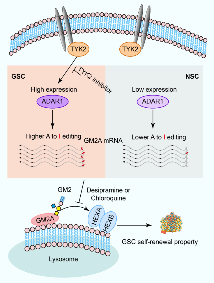
      Bookmark
          There is no author summary for this article yet. Authors can add summaries to their articles on ScienceOpen to make them more accessible to a non-specialist audience.

          Abstract

          Glioblastoma (GBM) is the most common and lethal primary malignant brain tumor, containing GBM stem cells (GSCs) that contribute to therapeutic resistance and relapse. Exposing potential GSC vulnerabilities may provide therapeutic strategies against GBM. Here, we interrogated the role of adenosine-to-inosine (A-to-I) RNA editing mediated by adenosine deaminase acting on RNA 1 (ADAR1) in GSCs and found that both ADAR1 and global RNA editomes were elevated in GSCs compared with normal neural stem cells. ADAR1 inactivation or blocking of the upstream JAK/STAT pathway through TYK2 inhibition impaired GSC self-renewal and stemness. Downstream of ADAR1, RNA editing of the 3′-UTR of GM2A, a key ganglioside catabolism activator, proved to be critical, as interference with ganglioside catabolism and disruption of ADAR1 showed a similar functional impact on GSCs. These findings reveal that RNA editing links ganglioside catabolism to GSC self-renewal and stemness, exposing a potential vulnerability of GBM for therapeutic intervention.

          Abstract

          Related collections

          Most cited references97

          • Record: found
          • Abstract: found
          • Article: found
          Is Open Access

          Moderated estimation of fold change and dispersion for RNA-seq data with DESeq2

          In comparative high-throughput sequencing assays, a fundamental task is the analysis of count data, such as read counts per gene in RNA-seq, for evidence of systematic changes across experimental conditions. Small replicate numbers, discreteness, large dynamic range and the presence of outliers require a suitable statistical approach. We present DESeq2, a method for differential analysis of count data, using shrinkage estimation for dispersions and fold changes to improve stability and interpretability of estimates. This enables a more quantitative analysis focused on the strength rather than the mere presence of differential expression. The DESeq2 package is available at http://www.bioconductor.org/packages/release/bioc/html/DESeq2.html. Electronic supplementary material The online version of this article (doi:10.1186/s13059-014-0550-8) contains supplementary material, which is available to authorized users.
            Bookmark
            • Record: found
            • Abstract: found
            • Article: found
            Is Open Access

            Trimmomatic: a flexible trimmer for Illumina sequence data

            Motivation: Although many next-generation sequencing (NGS) read preprocessing tools already existed, we could not find any tool or combination of tools that met our requirements in terms of flexibility, correct handling of paired-end data and high performance. We have developed Trimmomatic as a more flexible and efficient preprocessing tool, which could correctly handle paired-end data. Results: The value of NGS read preprocessing is demonstrated for both reference-based and reference-free tasks. Trimmomatic is shown to produce output that is at least competitive with, and in many cases superior to, that produced by other tools, in all scenarios tested. Availability and implementation: Trimmomatic is licensed under GPL V3. It is cross-platform (Java 1.5+ required) and available at http://www.usadellab.org/cms/index.php?page=trimmomatic Contact: usadel@bio1.rwth-aachen.de Supplementary information: Supplementary data are available at Bioinformatics online.
              Bookmark
              • Record: found
              • Abstract: found
              • Article: not found

              STAR: ultrafast universal RNA-seq aligner.

              Accurate alignment of high-throughput RNA-seq data is a challenging and yet unsolved problem because of the non-contiguous transcript structure, relatively short read lengths and constantly increasing throughput of the sequencing technologies. Currently available RNA-seq aligners suffer from high mapping error rates, low mapping speed, read length limitation and mapping biases. To align our large (>80 billon reads) ENCODE Transcriptome RNA-seq dataset, we developed the Spliced Transcripts Alignment to a Reference (STAR) software based on a previously undescribed RNA-seq alignment algorithm that uses sequential maximum mappable seed search in uncompressed suffix arrays followed by seed clustering and stitching procedure. STAR outperforms other aligners by a factor of >50 in mapping speed, aligning to the human genome 550 million 2 × 76 bp paired-end reads per hour on a modest 12-core server, while at the same time improving alignment sensitivity and precision. In addition to unbiased de novo detection of canonical junctions, STAR can discover non-canonical splices and chimeric (fusion) transcripts, and is also capable of mapping full-length RNA sequences. Using Roche 454 sequencing of reverse transcription polymerase chain reaction amplicons, we experimentally validated 1960 novel intergenic splice junctions with an 80-90% success rate, corroborating the high precision of the STAR mapping strategy. STAR is implemented as a standalone C++ code. STAR is free open source software distributed under GPLv3 license and can be downloaded from http://code.google.com/p/rna-star/.
                Bookmark

                Author and article information

                Contributors
                Journal
                J Clin Invest
                J Clin Invest
                J Clin Invest
                The Journal of Clinical Investigation
                American Society for Clinical Investigation
                0021-9738
                1558-8238
                15 March 2022
                15 March 2022
                15 March 2022
                15 March 2022
                : 132
                : 6
                : e143397
                Affiliations
                [1 ]Division of Regenerative Medicine, Department of Medicine, UCSD, La Jolla, California, USA.
                [2 ]Sanford Consortium for Regenerative Medicine, La Jolla, California, USA.
                [3 ]Department of Cellular and Molecular Medicine, UCSD, La Jolla, California, USA.
                [4 ]UPMC Hillman Cancer Center, Pittsburgh, Pennsylvania, USA.
                [5 ]Medical Scientist Training Program,
                [6 ]Cleveland Clinic Lerner College of Medicine, and
                [7 ]Department of Pathology, Case Western Reserve University, Cleveland, Ohio, USA.
                [8 ]Department of Neurology, University of Pittsburgh, Pittsburgh, Pennsylvania, USA.
                Author notes
                Address correspondence to: Jeremy N. Rich, UPMC Cancer Pavilion, Room 549, 5150 Centre Avenue, Pittsburgh, Pennsylvania 15232, USA. Phone: 412.623.3364; Email: drjeremyrich@ 123456gmail.com . Or to: Xiang-Dong Fu, Department of Cellular and Molecular Medicine, University of California, San Diego, 9500 Gilman Drive #0651, La Jolla, California 92093, USA. Phone: 858.534.4937; Email: xdfu@ 123456ucsd.edu .

                Authorship note: LJ and YH contributed equally to this work.

                Author information
                http://orcid.org/0000-0002-4082-7200
                http://orcid.org/0000-0002-7189-3243
                http://orcid.org/0000-0001-7103-8004
                http://orcid.org/0000-0002-5385-948X
                http://orcid.org/0000-0001-5499-8732
                http://orcid.org/0000-0001-7845-5302
                Article
                143397
                10.1172/JCI143397
                8920333
                35133980
                9c536ba5-8e73-420d-b606-b09196ec0d16
                © 2022 Li Jiang et al.

                This work is licensed under the Creative Commons Attribution 4.0 International License. To view a copy of this license, visit http://creativecommons.org/licenses/by/4.0/.

                History
                : 19 August 2020
                : 3 February 2022
                Funding
                Funded by: National Cancer Institute, https://doi.org/10.13039/100000054;
                Award ID: CA217066,CA217065,CA238662,CA197718,CA154130,CA169117,CA171652
                Funded by: National Institute of General Medical Sciences, https://doi.org/10.13039/100000057;
                Award ID: GM04936,GM052872
                Funded by: National Human Genome Research Institute, https://doi.org/10.13039/100000051;
                Award ID: HG004659
                Funded by: National Institute of Neurological Disorders and Stroke, https://doi.org/10.13039/100000065;
                Award ID: NS087913,NS089272,NS103434
                Funded by: National Cancer Institute, https://doi.org/10.13039/100000054;
                Award ID: CA047904
                Categories
                Research Article

                oncology,stem cells,brain cancer,human stem cells
                oncology, stem cells, brain cancer, human stem cells

                Comments

                Comment on this article