3
views
0
recommends
+1 Recommend
0 collections
    0
    shares
      • Record: found
      • Abstract: found
      • Article: found
      Is Open Access

      Willin/FRMD6 Influences Mechanical Phenotype and Neuronal Differentiation in Mammalian Cells by Regulating ERK1/2 Activity

      research-article

      Read this article at

      Bookmark
          There is no author summary for this article yet. Authors can add summaries to their articles on ScienceOpen to make them more accessible to a non-specialist audience.

          Abstract

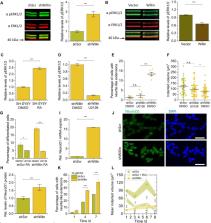
          Willin/FRMD6 is part of a family of proteins with a 4.1 ezrin-radixin-moesin (FERM) domain. It has been identified as an upstream activator of the Hippo pathway and, when aberrant in its expression, is associated with human diseases and disorders. Even though Willin/FRMD6 was originally discovered in the rat sciatic nerve, most studies have focused on its functional roles in cells outside of the nervous system, where Willin/FRMD6 is involved in the formation of apical junctional cell-cell complexes and in regulating cell migration. Here, we investigate the biochemical and biophysical role of Willin/FRMD6 in neuronal cells, employing the commonly used SH-SY5Y neuronal model cell system and combining biochemical measurements with Elastic Resonator Interference Stress Micropscopy (ERISM). We present the first direct evidence that Willin/FRMD6 expression influences both the cell mechanical phenotype and neuronal differentiation. By investigating cells with increased and decreased Willin/FRMD6 expression levels, we show that Willin/FRMD6 not only affects proliferation and migration capacity of cells but also leads to changes in cell morphology and an enhanced formation of neurite-like membrane extensions. These changes were accompanied by alterations of biophysical parameters such as cell force, the organization of actin stress fibers and the formation of focal adhesions. At the biochemical level, changes in Willin/FRMD6 expression inversely affected the activity of the extracellular signal-regulated kinases (ERK) pathway and downstream transcriptional factor NeuroD1, which seems to prime SH-SY5Y cells for retinoic acid (RA)-induced neuronal differentiation.

          Related collections

          Most cited references33

          • Record: found
          • Abstract: found
          • Article: not found

          Identification of a novel inhibitor of mitogen-activated protein kinase kinase.

          The compound U0126 (1,4-diamino-2,3-dicyano-1, 4-bis[2-aminophenylthio]butadiene) was identified as an inhibitor of AP-1 transactivation in a cell-based reporter assay. U0126 was also shown to inhibit endogenous promoters containing AP-1 response elements but did not affect genes lacking an AP-1 response element in their promoters. These effects of U0126 result from direct inhibition of the mitogen-activated protein kinase kinase family members, MEK-1 and MEK-2. Inhibition is selective for MEK-1 and -2, as U0126 shows little, if any, effect on the kinase activities of protein kinase C, Abl, Raf, MEKK, ERK, JNK, MKK-3, MKK-4/SEK, MKK-6, Cdk2, or Cdk4. Comparative kinetic analysis of U0126 and the MEK inhibitor PD098059 (Dudley, D. T., Pang, L., Decker, S. J., Bridges, A. J., and Saltiel, A. R. (1995) Proc. Natl. Acad. Sci U. S. A. 92, 7686-7689) demonstrates that U0126 and PD098059 are noncompetitive inhibitors with respect to both MEK substrates, ATP and ERK. We further demonstrate that the two compounds bind to deltaN3-S218E/S222D MEK in a mutually exclusive fashion, suggesting that they may share a common or overlapping binding site(s). Quantitative evaluation of the steady state kinetics of MEK inhibition by these compounds reveals that U0126 has approximately 100-fold higher affinity for deltaN3-S218E/S222D MEK than does PD098059. We further tested the effects of these compounds on the activity of wild type MEK isolated after activation from stimulated cells. Surprisingly, we observe a significant diminution in affinity of both compounds for wild type MEK as compared with the deltaN3-S218E/S222D mutant enzyme. These results suggest that the affinity of both compounds is mediated by subtle conformational differences between the two activated MEK forms. The MEK affinity of U0126, its selectivity for MEK over other kinases, and its cellular efficacy suggest that this compound will serve as a powerful tool for in vitro and cellular investigations of mitogen-activated protein kinase-mediated signal transduction.
            Bookmark
            • Record: found
            • Abstract: found
            • Article: not found

            YAP/TAZ upstream signals and downstream responses

            Cell behavior is strongly influenced by physical, mechanical contacts between cells and their extracellular matrix. We review how the transcriptional regulators YAP/TAZ integrate mechanical cues with the response to soluble signals and metabolic pathways to control multiple aspects of cell behavior, including proliferation, cell plasticity and stemness essential for tissue regeneration. Corruption of cell-environment interplay leads to aberrant YAP/TAZ activation that is instrumental for multiple diseases, including cancer.
              Bookmark
              • Record: found
              • Abstract: found
              • Article: not found

              YAP-dependent induction of amphiregulin identifies a non-cell-autonomous component of the Hippo pathway.

              The Hippo signalling pathway regulates cellular proliferation and survival, thus has profound effects on normal cell fate and tumorigenesis. The pivotal effector of this pathway is YAP (yes-associated protein), a transcriptional co-activator amplified in mouse and human cancers, where it promotes epithelial to mesenchymal transition (EMT) and malignant transformation. So far, studies of YAP target genes have focused on cell-autonomous mediators; here we show that YAP-expressing MCF10A breast epithelial cells enhance the proliferation of neighbouring untransfected cells, implicating a non-cell-autonomous mechanism. We identify the gene for the epidermal growth factor receptor (EGFR) ligand amphiregulin (AREG) as a transcriptional target of YAP, whose induction contributes to YAP-mediated cell proliferation and migration, but not EMT. Knockdown of AREG or addition of an EGFR kinase inhibitor abrogates the proliferative effects of YAP expression. Suppression of the negative YAP regulators LATS1 and 2 (large tumour suppressor 1 and 2) is sufficient to induce AREG expression, consistent with physiological regulation of AREG by the Hippo pathway. Genetic interaction between the Drosophila YAP orthologue Yorkie and Egfr signalling components supports the link between these two highly conserved signalling pathways. Thus, YAP-dependent secretion of AREG indicates that activation of EGFR signalling is an important non-cell-autonomous effector of the Hippo pathway, which has implications for the regulation of both physiological and malignant cell proliferation.
                Bookmark

                Author and article information

                Contributors
                Journal
                Front Cell Neurosci
                Front Cell Neurosci
                Front. Cell. Neurosci.
                Frontiers in Cellular Neuroscience
                Frontiers Media S.A.
                1662-5102
                04 September 2020
                2020
                : 14
                : 552213
                Affiliations
                [1] 1Centre of Biophotonics and SUPA, School of Physics and Astronomy, University of St Andrews , St Andrews, United Kingdom
                [2] 2Centre for Nanobiophotonics, Department of Chemistry, University of Cologne , Cologne, Germany
                [3] 3Centre of Biophotonics, School of Biology, University of St Andrews , St Andrews, United Kingdom
                [4] 4Department of Biochemistry, School of Medicine, Boston University , Boston, MA, United States
                [5] 5Department of Physics, College of Science, Yonsei University , Seoul, South Korea
                Author notes

                Edited by: Tommaso Pizzorusso, University of Florence, Italy

                Reviewed by: Vincenza Rita Lo Vasco, Independent researcher, Florence, Italy; Danuta Jantas, Polish Academy of Sciences (IF PAS), Poland

                *Correspondence: Malte C. Gather mcg6@ 123456st-andrews.ac.uk Frank J. Gunn-Moore fjg1@ 123456st-andrews.ac.uk

                These authors have contributed equally to this work

                Specialty section: This article was submitted to Cellular Neurophysiology, a section of the journal Frontiers in Cellular Neuroscience

                Article
                10.3389/fncel.2020.552213
                7498650
                9be9b0d3-9834-4624-bad8-94a5fc6339ce
                Copyright © 2020 Kronenberg, Tilston-Lunel, Thompson, Chen, Yu, Dholakia, Gather and Gunn-Moore.

                This is an open-access article distributed under the terms of the Creative Commons Attribution License (CC BY). The use, distribution or reproduction in other forums is permitted, provided the original author(s) and the copyright owner(s) are credited and that the original publication in this journal is cited, in accordance with accepted academic practice. No use, distribution or reproduction is permitted which does not comply with these terms.

                History
                : 15 April 2020
                : 17 August 2020
                Page count
                Figures: 4, Tables: 0, Equations: 0, References: 33, Pages: 11, Words: 8026
                Funding
                Funded by: Engineering and Physical Sciences Research Council 10.13039/501100000266
                Funded by: Biotechnology and Biological Sciences Research Council 10.13039/501100000268
                Categories
                Cellular Neuroscience
                Brief Research Report

                Neurosciences
                willin/frmd6,neuronal differentiation,erk1/2,cell mechanics,actin,focal adhesion,taz,cell force measurement

                Comments

                Comment on this article