6
views
0
recommends
+1 Recommend
0 collections
    0
    shares
      • Record: found
      • Abstract: found
      • Article: found
      Is Open Access

      Prostaglandin E 2 Dilates Intracerebral Arterioles When Applied to Capillaries: Implications for Small Vessel Diseases

      research-article

      Read this article at

      Bookmark
          There is no author summary for this article yet. Authors can add summaries to their articles on ScienceOpen to make them more accessible to a non-specialist audience.

          Abstract

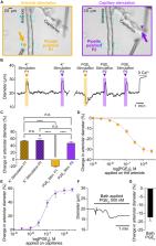
          Prostaglandin E 2 (PGE 2) has been widely proposed to mediate neurovascular coupling by dilating brain parenchymal arterioles through activation of prostanoid EP4 receptors. However, our previous report that direct application of PGE 2 induces an EP1-mediated constriction strongly argues against its direct action on arterioles during neurovascular coupling, the mechanisms sustaining functional hyperemia. Recent advances have highlighted the role of capillaries in sensing neuronal activity and propagating vasodilatory signals to the upstream penetrating parenchymal arteriole. Here, we examined the effect of capillary stimulation with PGE 2 on upstream arteriolar diameter using an ex vivo capillary-parenchymal arteriole preparation and in vivo cerebral blood flow measurements with two-photon laser-scanning microscopy. We found that PGE 2 caused upstream arteriolar dilation when applied onto capillaries with an EC 50 of 70 nM. The response was inhibited by EP1 receptor antagonist and was greatly reduced, but not abolished, by blocking the strong inward-rectifier K + channel. We further observed a blunted dilatory response to capillary stimulation with PGE 2 in a genetic mouse model of cerebral small vessel disease with impaired functional hyperemia. This evidence casts previous findings in a different light, indicating that capillaries are the locus of PGE 2 action to induce upstream arteriolar dilation in the control of brain blood flow, thereby providing a paradigm-shifting view that nonetheless remains coherent with the broad contours of a substantial body of existing literature.

          Related collections

          Most cited references51

          • Record: found
          • Abstract: found
          • Article: not found

          Cerebral small vessel disease: from pathogenesis and clinical characteristics to therapeutic challenges.

          The term cerebral small vessel disease refers to a group of pathological processes with various aetiologies that affect the small arteries, arterioles, venules, and capillaries of the brain. Age-related and hypertension-related small vessel diseases and cerebral amyloid angiopathy are the most common forms. The consequences of small vessel disease on the brain parenchyma are mainly lesions located in the subcortical structures such as lacunar infarcts, white matter lesions, large haemorrhages, and microbleeds. Because lacunar infarcts and white matter lesions are easily detected by neuroimaging, whereas small vessels are not, the term small vessel disease is frequently used to describe the parenchyma lesions rather than the underlying small vessel alterations. This classification, however, restricts the definition of small vessel disease to ischaemic lesions and might be misleading. Small vessel disease has an important role in cerebrovascular disease and is a leading cause of cognitive decline and functional loss in the elderly. Small vessel disease should be a main target for preventive and treatment strategies, but all types of presentation and complications should be taken into account. Copyright 2010 Elsevier Ltd. All rights reserved.
            Bookmark
            • Record: found
            • Abstract: found
            • Article: not found

            A molecular atlas of cell types and zonation in the brain vasculature

            Cerebrovascular disease is the third most common cause of death in developed countries, but our understanding of the cells that compose the cerebral vasculature is limited. Here, using vascular single-cell transcriptomics, we provide molecular definitions for the principal types of blood vascular and vessel-associated cells in the adult mouse brain. We uncover the transcriptional basis of the gradual phenotypic change (zonation) along the arteriovenous axis and reveal unexpected cell type differences: a seamless continuum for endothelial cells versus a punctuated continuum for mural cells. We also provide insight into pericyte organotypicity and define a population of perivascular fibroblast-like cells that are present on all vessel types except capillaries. Our work illustrates the power of single-cell transcriptomics to decode the higher organizational principles of a tissue and may provide the initial chapter in a molecular encyclopaedia of the mammalian vasculature.
              Bookmark
              • Record: found
              • Abstract: found
              • Article: not found

              Small vessel disease: mechanisms and clinical implications

              Small vessel disease is a disorder of cerebral microvessels that causes white matter hyperintensities and several other common abnormalities (eg, recent small subcortical infarcts and lacunes) seen on brain imaging. Despite being a common cause of stroke and vascular dementia, the underlying pathogenesis is poorly understood. Research in humans has identified several manifestations of cerebral microvessel endothelial dysfunction including blood-brain barrier dysfunction, impaired vasodilation, vessel stiffening, dysfunctional blood flow and interstitial fluid drainage, white matter rarefaction, ischaemia, inflammation, myelin damage, and secondary neurodegeneration. These brain abnormalities are more dynamic and widespread than previously thought. Relationships between lesions and symptoms are highly variable but poorly understood. Major challenges are the determination of which vascular dysfunctions are most important in pathogenesis, which abnormalities are reversible, and why lesion progression and symptomatology are so variable. This knowledge will help to identify potential targets for intervention and improve risk prediction for individuals with small vessel disease.
                Bookmark

                Author and article information

                Contributors
                Journal
                Front Aging Neurosci
                Front Aging Neurosci
                Front. Aging Neurosci.
                Frontiers in Aging Neuroscience
                Frontiers Media S.A.
                1663-4365
                13 August 2021
                2021
                : 13
                : 695965
                Affiliations
                [1] 1Department of Anesthesiology, University of Colorado Anschutz Medical Campus , Aurora, CO, United States
                [2] 2Department of Physiology, School of Medicine, University of Maryland , Baltimore, Baltimore, MD, United States
                [3] 3INSERM, UMR 1266, GHU Paris Psychiatrie et Neurosciences, Institute of Psychiatry and Neuroscience of Paris, University of Paris , Paris, France
                [4] 4Department of Pharmacology, Larner College of Medicine, University of Vermont , Burlington, VT, United States
                [5] 5Department of Pharmacology, University of Colorado Anschutz Medical Campus , Aurora, CO, United States
                Author notes

                Edited by: Anusha Mishra, Oregon Health and Science University, United States

                Reviewed by: Cam Ha Tran, University of Nevada, Reno, United States; George C. Wellman, University of Vermont, United States; Eric Newman, University of Minnesota, United States

                *Correspondence: Fabrice Dabertrand, Fabrice.Dabertrand@ 123456CUAnschutz.edu
                Article
                10.3389/fnagi.2021.695965
                8414797
                34483880
                9be87c72-1989-48c4-8ee1-666e71f19811
                Copyright © 2021 Rosehart, Longden, Weir, Fontaine, Joutel and Dabertrand.

                This is an open-access article distributed under the terms of the Creative Commons Attribution License (CC BY). The use, distribution or reproduction in other forums is permitted, provided the original author(s) and the copyright owner(s) are credited and that the original publication in this journal is cited, in accordance with accepted academic practice. No use, distribution or reproduction is permitted which does not comply with these terms.

                History
                : 15 April 2021
                : 15 June 2021
                Page count
                Figures: 6, Tables: 0, Equations: 0, References: 51, Pages: 11, Words: 0
                Funding
                Funded by: National Heart, Lung, and Blood Institute 10.13039/100000050
                Award ID: R01HL136636
                Funded by: Center for Women’s Health Research, University of Colorado 10.13039/100012876
                Categories
                Neuroscience
                Original Research

                Neurosciences
                functional hyperemia,cerebral small vessel diseases,cadasil,microcirculation,neurovascular coupling,potassium channel,prostaglandin e2,epidermal growth factor receptor

                Comments

                Comment on this article

                scite_
                0
                0
                0
                0
                Smart Citations
                0
                0
                0
                0
                Citing PublicationsSupportingMentioningContrasting
                View Citations

                See how this article has been cited at scite.ai

                scite shows how a scientific paper has been cited by providing the context of the citation, a classification describing whether it supports, mentions, or contrasts the cited claim, and a label indicating in which section the citation was made.

                Similar content158

                Cited by6

                Most referenced authors459